Asian Spine J Search

CLOSE


Asian Spine J > Volume 18(1); 2024 > Article
Fong, Lim, Tan, and Hey: A Comparison between Structural Allografts and Polyetheretherketone Interbody Spacers Used in Anterior Cervical Discectomy and Fusion: A Systematic Review and Meta-analysis

Abstract

Among interbody implants used during anterior cervical discectomy and fusion (ACDF), structural allografts and polyetheretherketone (PEEK) are the most used spacers. Currently, no consensus has been established regarding the superiority of either implant, with US surgeons preferring structural allografts, whereas UK surgeons preferring PEEK. The purpose of this systematic review (level of evidence, 4) was to compare postoperative and patient-reported outcomes between the use of structural allografts PEEK interbody spacers during ACDF. Five electronic databases (PubMed, Embase, Scopus, Web of Science, and Cochrane) were searched for articles comparing the usage of structural allograft and PEEK interbody spacers during ACDF procedures from inception to April 10, 2023. The searches were conducted using the keywords “Spine,” “Allograft,” and “PEEK” and were performed in accordance with the Preferred Reporting Items for Systematic Reviews and Meta-analysis guidelines. Subsequent quality and sensitivity analyses were performed on the included studies. Nine studies involving 1,074 patients were included. Compared with the PEEK group, the structural allograft group had comparable rates of postoperative pseudoarthrosis (p=0.58). However, when stratified according to the number of levels treated, the 3-level ACDF PEEK group was 3.45 times more likely to have postoperative pseudoarthrosis than the structural allograft group (p=0.01). Subsequent postoperative outcomes (rate of subsidence and change in the preoperative and postoperative segmental disc heights) were comparable between the PEEK and structural allograft groups. Patient-reported outcomes (Visual Analog Scale [VAS] of neck pain and Neck Disability Index [NDI]) were comparable. This study showed that for 3-level ACDFs, the use of structural allografts may confer higher fusion rates. However, VAS neck pain, NDI, and subsidence rates were comparable between structural allografts and PEEK cages. In addition, no significant difference in pseudoarthrosis rates was found between PEEK cages and structural allografts in patients undergoing 1- and 2-level ACDFs.

Introduction

Anterior cervical discectomy and fusion (ACDF) is a well-established treatment for cervical myelopathy and radiculopathy [14]. It was first described by Smith and Robinson [4] in 1958, who advocated the use of a structural iliac crest autograft to promote interbody fusion. However, the use of harvested iliac crest grafts has complications and is associated with donor-site morbidities such as pain and surgical site infection [5]. As a result, various alternatives such as interbody cages and structural allografts were developed to mitigate these complications [6].
Among interbody implants, structural allografts and polyetheretherketone (PEEK) are the most commonly used spacers. In a survey comprising 599 AO spine members, 92% utilized structural allograft or PEEK interbody cages during ACDF [7]. However, no consensus has been established regarding the superiority of either implant, with US surgeons preferring structural allografts, where UK surgeons preferring PEEK cages [8].
Previous systematic reviews [9] and meta-analysis [10] have attempted to compare PEEK cages with structural allografts as interbody spacers, and both cervical and lumbar data were combined because of insufficient data. Thus, this review aimed to compare the postoperative clinical and radiological outcomes of PEEK interbody cages with structural allograft interbody implants in ACDF. To the author’s knowledge, this is the first meta-analysis that seeks to compare PEEK cages against structural allografts in ACDF procedures.

Methods

1. Search strategy

The study was conducted following the Preferred Reporting Items for Systematic Reviews and Meta-analysis statement [11]. An electronic database search of PubMed, Embase, Scopus, Web of Science, and Cochrane Central Register of Controlled Trials was performed from inception to April 10, 2023 using keywords relating to spine (spinal OR spine* OR vertebra* OR vertebrae* OR cervical OR “cervical spine*”), ACDF (surgery OR surgeries OR operation* OR surgical OR procedure* OR “cervical fusion” OR “interbody fusion” OR interbody* OR ACDF OR “Anterior Cervical Discectomy and Fusion” OR discectomy* OR diskectomy*), allograft (allograft* OR homograft* OR allotransplant* OR “structural allograft” OR allogenic OR allogeneic OR homologous OR homogenous OR bone transplant* OR bone graft*), and PEEK (PEEK OR polyetheretherketone OR Cage* OR polyetherketone).

2. Data assessment and inclusion and exclusion criteria

Abstracts were screened to remove duplicates and selected based on the predetermined inclusion criteria (Table 1). The full texts of the remaining studies were further analyzed. The reference lists of the included articles were manually searched to identify further studies for analysis. Any discrepancies were resolved by achieving a consensus with a third author (T.J.H.).
Studies comparing the use of structural allografts with PEEK interbody cages for ACDF were selected. The Newcastle–Ottawa Quality Assessment Scale was used to assess the quality of non-randomized studies in meta-analyses [12]. All nine included studies were of good quality according to the Newcastle–Ottawa Scale, with a range of 7–9 points (Supplement 1).

3. Data collection

Data from the included studies were extracted independently by F.F.J.Y. and L.C.Y. using a standardized protocol and reporting form. Study characteristics (year of study, study design, follow-up duration, and patient demographics) and operative outcomes (pseudoarthrosis, subsidence rates, and segmental disc height) were extracted. The following patient-reported outcomes at baseline and last follow-up were also extracted: Visual Analog Scale (VAS) for neck pain and Neck Disability Index (NDI). When means and standard deviations were unavailable, data were converted using previously established models by Wan et al. [13]. The corresponding authors were not contacted because no data were missing during extraction. The following definitions for operative and patient-reported outcomes were used:
  • (1) Pseudoarthrosis was defined as (a) no bridging bone seen across the intervertebral space, (b) radiolucent line at the graft vertebral body interface, and (c) motion >1 mm between adjacent spinous processes as seen on flexion–extension cervical spine radiographs in all of the studies [1418]. Wang et al. [14] further included patients who had a halo appearing around the screws and/or implant failure that could be visualized on radiographs into their definition of pseudoarthrosis.

  • (2) Subsidence was defined as a radiologic decrease in the intervertebral height of ≥2 mm between the immediate postoperative and final postoperative radiographs [7,17,19,20]. All studies that reported subsidence adopted this definition.

  • (3) The segmental disc height was defined as either the average of the anterior and posterior intervertebral height [21] or the midpoint of the upper endplate of the vertebral body to the midpoint of the lower endplate of the vertebral body [14,17].

  • (4) VAS neck pain was reported on a scale of 0–10, where 0 was defined as “no pain,” whereas 10 as “worst pain ever” [22].

  • (5) Patient disability was measured using NDI, with scores ranging from 0 to 50 points [23].

4. Statistical analysis

For each measured outcome, baseline variables such as age and sex were statistically compared the structural allograft group and the PEEK group. For these outcome variables, age and sex were not statistically significant between the two groups, indicating that these two variables were comparable at baseline (Table 2). The odds ratio for binary outcomes and the mean difference for continuous outcomes were extracted from the selected studies. Heterogeneity among studies was assessed using the I2 statistic, interpreted according to definitions provided by the Cochrane Handbook, where an I2 value of 0%–50% represents heterogeneity that might not be important and 50%–100% may represent heterogeneity [24]. Statistical significance was considered at p≤0.05. All statistical analyses were made assuming a two-sided test at 5% level of significance using the Review Manager ver. 5.3 (Revman; Cochrane, London, UK). Random-effects models were utilized in all meta-analyses.

Results

A systematic literature search using our search strategy yielded a total of 5,393 articles, with 2,110 remaining after duplicate removal. Moreover, 2,070 articles were excluded based on the title and abstract review. The remaining 40 articles underwent full-text review, of which nine articles were subsequently included in the meta-analysis (Fig. 1). Five articles were retrospective studies, three were prospective studies, and one was a randomized controlled trial. The study characteristics and clinical outcomes are summarized in Tables 3 and 4, respectively.
The study included a total of 1,074 patients who underwent ACDF instrumentation for spinal surgery. Of these patients, 667 underwent structural allograft fixation for ACDF, whereas 407 underwent PEEK cage fixation for ACDF. All nine studies included patients who had anterior cervical plating [7,1420,25]. In general, seven papers did not specify whether bone morphogenic protein (BMP) was utilized [7,15,1720,25], whereas the remaining two papers stated that none of their patients used BMP [14,16].

Study Characteristics

1. Incidence of pseudoarthrosis

The pooled numbers of patients who had postoperative pseudoarthrosis following instrumentation with structural allografts from five studies involving 445 patients and following instrumentation with PEEK cages from five studies involving 246 patients were 97 and 51, respectively.
In five studies involving 691 patients, the estimated pooled odds ratio of patients who had postoperative pseudoarthrosis between structural allografts and PEEK cages, was 1.33 (95% confidence interval [CI], 0.68–2.59; p=0.40; I2=35%) (Fig. 2). These findings suggest the lack of significant difference in the postoperative pseudoarthrosis rates between the structural allograft group and PEEK groups.
In a subgroup analysis of three studies comparing 266 patients who underwent 1-level ACDF, the pooled odds ratio of patients between the structural allograft and PEEK groups that had postoperative pseudoarthrosis was 0.65 (95% CI, 0.28–1.53; p=0.32; I2=0%) (Fig. 2). In a subgroup analysis of three studies involving 272 patients who underwent 2-level ACDF, the pooled odds ratio of patients between the structural allograft and PEEK groups that had postoperative pseudoarthrosis was 1.09 (95% CI, 0.35–3.42; p=0.88; I2=36%) (Fig. 2). In a subgroup analysis of three studies involving 153 patients who underwent 3-level ACDF, the pooled odds ratio of patients between the structural allograft and PEEK groups that had postoperative pseudoarthrosis was 3.45 (95% CI, 1.28–9.25; p=0.01; I2=0%) (Fig. 2).
These findings suggest that pseudoarthrosis rates were comparable between the structural allograft and PEEK groups at 1- and 2-level ACDFs. However, in 3-level ACDF, PEEK cages were 3.45 times significantly more likely to result in pseudoarthrosis than structural allografts.

2. Incidence of subsidence

In four studies involving 238 patients, the pooled number of patients who had postoperative subsidence following instrumentation with structural allografts was 59. In four studies involving 197 patients, the pooled number of patients who had postoperative subsidence following instrumentation with PEEK cages was 56.
From the four studies involving 435 patients, the estimated pooled odds ratio of patients in the structural allograft and PEEK group who had postoperative subsidence was 1.32 (95% CI, 0.74–2.33; p=0.34; I2=29%) (Fig. 3). These findings suggest that the subsidence rates were comparable between the structural allograft and PEEK groups.

3. Segmental disc height

In four studies involving 332 patients, the pooled change between the preoperative and postoperative segmental disc heights in the structural allograft group was 1.10±2.59 mm. In four studies involving 222 patients, the pooled change between the preoperative and postoperative segmental disc heights in the PEEK group was 1.71±2.86 mm. In four studies involving 554 patients, the standard difference in the means of the segmental disc height between the structural allograft and PEEK groups was analyzed, giving a pooled estimate of −0.03 (95% CI, −0.42 to 0.36; p=0.89; I2=38%) (Fig. 4).
In a subgroup analysis of three studies comparing 213 patients who underwent 1-level ACDF, the pooled change between the preoperative and postoperative segmental disc heights between the structural allograft and PEEK groups was 0.02 (95% CI, −0.29 to 0.33; p=0.89; I2=0%) (Fig. 4). In a subgroup analysis of four studies involving 259 patients who underwent 2-level ACDF, the pooled change between the preoperative and postoperative segmental disc heights between the structural allograft and PEEK groups was −0.21 (95% CI, −1.20 to 0.79; p=0.69; I2=63%) (Fig. 4). In a subgroup analysis of three studies involving 82 patients who underwent 3-level ACDF, the pooled change between the preoperative and postoperative segmental disc heights in the structural allograft and PEEK groups was −1.57 (95% CI, −6.01 to 2.88; p=0.49; I2=73%) (Fig. 4). These findings suggest that the difference in the preoperative and postoperative segmental disc heights was comparable between the structural allograft and PEEK groups.

4. VAS neck pain

In four studies involving 311 patients, the pooled change between preoperative and postoperative VAS neck pain scores for the structural allograft group was 2.73±3.88 mm. In four studies involving 173 patients, the pooled change between preoperative and postoperative VAS neck pain scores in the PEEK cage group was 3.19±3.56 mm. In four studies involving 484 patients, the standard difference in the mean VAS neck pain score between the structural allograft and PEEK groups, was analyzed, giving a pooled estimate of −0.32 (95% CI, −0.72 to 0.08; p=0.12; I2=0%) (Fig. 5). These findings suggest that the change in preoperative and postoperative VAS neck pain scores was comparable between the structural allograft and PEEK groups.

5. NDI

In four studies involving 311 patients, the pooled change between the preoperative and postoperative NDI scores in the structural allograft group was 15.02±25.33 mm. In four studies involving 173 patients, the pooled change between the preoperative and postoperative NDI scores in the PEEK group was 20.30±21.31 mm. In four studies involving 484 patients, the standard difference in the mean NDI score between the structural allograft and PEEK groups was analyzed, giving a pooled estimate of −0.45 (95% CI, −1.82 to 0.93; p=0.52; I2=0%) (Fig. 6). These findings suggest that the change in the preoperative and postoperative NDI scores was comparable between the structural allograft and PEEK groups.

Discussion

PEEK cages are notable for their biocompatibility and corrosion-resistant and radiolucent properties, which enable clear visualization of fusion on radiography [26,27]. However, given its biologically inert property, PEEK cages do not provide osteoinduction, which may increase the risk of pseudoarthrosis and graft subsidence [2729]. On the contrary, structural allografts have the advantages of better osteoinduction but are associated with possible disease transmission [30] and are more radio-opaque than PEEK, which can make assessment of postoperative fusion challenging.
Pseudarthrosis is not an uncommon complication in ACDF and can be detrimental when it leads to inappropriate segmental motion. Patients may experience postoperative pain, neurological compromise (such as radiculopathy and myelopathy), and hardware failure and may require revision surgery [31]. Thus, this study found that in 1- and 2-level ACDFs, structural allografts and PEEK cages have comparable pseudarthrosis rates. However, in 3-level ACDF, the PEEK group was 3.45 times more likely to have pseudoarthrosis than the structural allograft group. A possible explanation could be PEEK cages’ inherent bioinert property that hinders their ability to achieve adequate osteointegration, subsequently reducing fusion rates [27]. In vitro studies observed that when mesenchymal cells are cultured on PEEK materials, they had significantly high levels of interleukin-1β, which is associated with the formation of fibrous tissues. Furthermore, these mesenchymal cells did not express known markers of bone formation [32]. An in vivo sheep model corroborated these findings, observing that PEEK cages were surrounded by fibrous tissue preventing osteointegration, increasing the risk of pseudoarthrosis [26]. In previous studies, multilevel ACDF was associated with increased rates of pseudoarthrosis compared with 1-level ACDF because of increased bone interfaces required for fusion and altered biomechanics [3335]. In 1- and 2-level ACDFs, more remaining mobile segments allow for residual cervical spine movement. The strain at the target fusion levels is correspondingly lower, which provides a more conducive environment for fusion. However, in 3-level ACDFs, where six vertebral endplates are prepared for fusion, fewer mobile segments remained. During the arc of motion, the strain experienced in the three immobilized segments is higher, leading to a propensity for pseudarthrosis. Furthermore, with high levels of interleukin-1β induced by the PEEK material, the mesenchymal cells across the six endplates can be biologically “overtaxed,” which results in pseudarthrosis. Thus, these could explain the difference in pseudoarthrosis rates only being observed in 3-level ACDFs. For 3-level ACDFs, one should consider the use of structural allografts instead of PEEK cages to reduce the postoperative risk of pseudarthrosis.
Implant subsidence is not uncommon and occurs in up to 96% postoperatively [3641]. It is hypothesized to occur in an implant that has a higher Young’s modulus than the native vertebral body [42]. Uncontrolled subsidence may lead to decreased intervertebral disc height and subsequent foraminal narrowing with symptom recurrence. This study found that the incidence and radiological degree of subsidence were comparable between the structural allograft and PEEK groups. When compared with other materials, PEEK cages have significantly lower elastic modulus, resulting in reduced stress shielding, increased bone growth, and hence lower subsidence rates [43]. For instance, titanium cages have significantly higher subsidence rates (20.5%) than PEEK cages (14.3%) [4446]. Furthermore, carbon fiber-based cages had a higher subsidence rate (25.2%) than PEEK cages (22.8%) [47]. We hypothesize that the comparable subsidence rates between structural allograft and PEEK cages are due to the minimal difference in Young’s modulus between the two materials [4851]. The elastic modulus of PEEK is approximately 8.3 GPa, which is similar to human cortical bone (17.7 GPa), and lower than that of titanium (116 GPa) [52]. Thus, this minute difference may not result in any clinical difference of subsidence.
In this study, no significant difference in NDI and VAS neck was found between structural allografts and PEEK cages. Patient-reported outcome scores are significantly poorer in patients with pseudoarthrosis requiring revision surgery [53]. In this study, no overall significant difference in fusion rates was found between PEEK cages and structural allografts when comparing all ACDF levels. Given that pseudoarthrosis rates were significantly higher in patients who underwent 3-level ACDF with PEEK cages, patient-related outcome scores may have been lower in patients who underwent PEEK cage insertion in the 3-level ACDF.
This study has several limitations. First, seven of the included articles were retrospective. To minimize the inherent bias of retrospective studies, the NOS scale was utilized to appraise the methodology of each included article. Articles that scored <7 were excluded. The final articles included had a mean score of 8.6 (range, 7–9). Second, some of the included studies have a minimum follow-up period of 1 year. Future studies with longer follow-ups are needed to corroborate our findings. Lastly, data on smoking status we insufficient for analysis, which has significantly increased the risk of pseudoarthrosis in spinal fusions [54].

Conclusions

This study showed that for 3-level ACDF, the use of structural allografts may confer higher fusion rates. However, neck pain VAS, NDI, and subsidence rates were comparable between structural allografts and PEEK cages. In addition, no significant difference in pseudoarthrosis rates was found between PEEK cages and structural allografts in patients undergoing 1- and 2-level ACDFs. Additional evidence of good-quality observational studies with a longer follow-up is needed to support our view of choosing structural allografts over PEEK cages.

Notes

Conflict of Interest

No potential conflict of interest relevant to this article was reported.

Author Contributions

Francis Fong Jia Yi and Lim Chee Yit contributed equally and are joint first authors. Francis Fong Jia Yi, Lim Chee Yit, and Tan Jun Hao contributed equally in the conceptualization of the study design and topic, data collection, data extraction, data analysis, and manuscript writing. Dennis Hey contributed in the manuscript writing.

Supplementary Materials

Supplementary materials can be available from https://doi.org/10.31616/2023.0128.
Supplement 1. Assessment of quality of studies included using the Newcastle Ottawa Scale.
asj-2023-0128-Supplementary-1.pdf

Fig. 1
Preferred Reporting Items for Systematic Reviews and Meta-Analyses (PRISMA) diagram. ACDF, anterior cervical discectomy and fusion.
asj-2023-0128f1.jpg
Fig. 2
Comparison of pseudoarthrosis rates. PEEK, polyetheretherketone; M-H, Mantel-Haenszel; CI, confidence interval; ACDF, anterior cervical discectomy and fusion; df, degrees of freedom.
asj-2023-0128f2.jpg
Fig. 3
Comparison of subsidence rates. PEEK, polyetheretherketone; M-H, Mantel-Haenszel; CI, confidence interval; df, degrees of freedom.
asj-2023-0128f3.jpg
Fig. 4
Comparison of postoperative changes in segmental disc height. SD, standard deviation; PEEK, polyetheretherketone; IV, inverse variance; CI, confidence interval; ACDF, anterior cervical discectomy and fusion; df, degrees of freedom.
asj-2023-0128f4.jpg
Fig. 5
Comparison of VAS Neck score. SD, standard deviation; PEEK, polyetheretherketone; IV, inverse variance; CI, confidence interval; df, degrees of freedom.
asj-2023-0128f5.jpg
Fig. 6
Comparison of NDI score. SD, standard deviation; PEEK, polyetheretherketone; IV, inverse variance; CI, confidence interval; df, degrees of freedom.
asj-2023-0128f6.jpg
Table 1
Inclusion and exclusion criteria
Inclusion Exclusion
  • - Comparative studies of structural allograft and PEEK following ACDF irrespective of the level of fusion

  • - Observational cohort or randomized control trial

  • - Minimum follow-up duration of 1 year

  • - Studies without a comparison group

  • - Biomechanical studies

  • - Cadaveric studies

  • - Animal studies

  • - In vitro studies

  • - Review articles, case reports, conference papers, and letters which do not contain original data

  • - Non-English language articles

  • - Studies that do not utilize anterior cervical plating

PEEK, polyetheretherketone; ACDF, anterior cervical discectomy and fusion.

Table 2
Baseline comparison between allograft and PEEK groups
Variable Allograft PEEK p-value
Pseudoarthrosis
 Age (yr) 55.91±11.68 56.53±11.10 0.60
 Male 166 75 0.81
Subsidence
 Age (yr) 55.24±10.68 54.20±10.02 0.30
 Male 125 79 0.12
Segmental disc height
 Age (yr) 54.37±10.53 54.79±11.36 0.68
 Male 162 94 0.36
VAS and NDI
 Age (yr) 54.96±10.65 56.87±10.80 0.06
 Male 151 76 0.56

Values are presented as mean±standard deviation or number. All p-values between baseline demographic data were statistically insignificant.

PEEK, polyetheretherketone; VAS, Visual Analog Scale; NDI, Neck Disability Index.

Table 3
Summary of the study characteristics
Study Study type Country Interbody devices used Mean age of patients (yr) Male Smoker BMI Levels of fusion FU duration (mo) Use of BMP
Yson et al. [7] (2017) Retrospective USA Total: 67; allograft: 19 (fibular allograft); PEEK: 48 Allograft: 54.4±13.3; PEEK: 51.1±10.8 Total: 28; allograft: 7; PEEK: 21 NR NR All 1-level >14 NR
Wang et al. [14] (2019) Retrospective USA Total: 168; allograft: 107 (unspecified); PEEK: 61 Allograft: 56±10.6; PEEK: 53.5±12.3 Total: 112; allograft: 75; PEEK: 37 Total: 59; allograft: 41; PEEK: 18 NR 1- Level: 96; 2-level: 104; 3-level: 64 >24 No patients received BMP
Yang et al. [25] (2019) Prospective China Total: 107; allograft: 58 (BioCage); PEEK: 49 Allograft: 50.3±12.3; PEEK: 50.6±11.8 Total: 59; allograft: 38; PEEK: 21 NR NR 1-Level: 80; 2-level: 27 >24 NR
Buyuk et al. [19] (2020) Retrospective USA Total: 209; allograft: 167 (unspecified); PEEK: 42 Allograft: 49.5±6.7; PEEK: 49.8±7.2 Total: 91; allograft: 69; PEEK: 22 Total: 43; allograft: 35; PEEK: 8 NR 1-Level: 84; 2-level: 125 >24 NR
Moo et al. [17] (2020) Prospective Singapore Total: 88; allograft: 53 (saline-packaged femoral or tibial cortical–cancellous allograft); PEEK: 35 (packed with local autografts from anterior osteophytes and shavings from burring the uncinate process) Allograft: 53.0±4.2; PEEK: 55.9±3.8 Total: 46; allograft: 27; PEEK: 19 Total: 10; allograft: 6; PEEK: 4 Allograft: 25.0±1.1; PEEK: 34.1±10.0 All 2-level >24 NR
Nchols et al. [18] (2020) Retrospective USA Total: 44; allograft: 31 (wedge iliac crest [17/31]; cortical cornerstone [14/31]); PEEK: 12; autograft: 1 Total: 57.0 Total: 22 Total: 6 current; 38 former or never Total: 27.8±5.29 All 3-level >24 NR
Ryu et al. [16] (2021) Prospective USA Total: 194; allograft: 154 (unspecified); PEEK: 40 Allograft: 56.7±10.7; PEEK: 61.8±8.9 Total: 83; allograft: 64; PEEK: 19 Total: 34; allograft: 30; PEEK: 4 NR 1-Level: 68; 2-level: 80; 3-level: 46 >12 No patients received BMP
Bergin et al. [15] (2021) Retrospective USA Total: 102; allograft: 32 (unspecified); PEEK: 70 Allograft: 53.8±13.9; PEEK: 54.1±12.1 NR NR Allograft: 29.2±6.13; PEEK: 30.0±7.33 All 1-level >12 NR
Villavicencio et al. (2022) RCT USA Total: 95; allograft: 46 (unspecified); PEEK: 49 Allograft: 57.3±11.5; PEEK: 59.8±11.6 Total: 39; allograft: 22; PEEK: 17 Total: 7; allograft: 4; PEEK: 3 NR 1-Level: 37; 2-level: 40; 3-level: 18 >24 NR

BMI, body mass index; FU, follow-up; BMP, bone morphogenic protein; PEEK, polyetheretherketone; NR, not reported; RCT, randomized controlled trial.

Table 4
Outcomes of included studies
Study Location of pseudoarthrosis Incidence of pseudoarthrosis Subsidence Segmental disc height (mm) VAS neck NDI
Yson et al. [7] (2017) NR NR Allograft: 9/32; PEEK: 25/85 NR NR NR
Wang et al. [14] (2019) NR 1 Level ACDF; allograft: 2/59; PEEK: 2/37
2 Level ACDF; allograft: 5/62; PEEK: 3/42
3 Level ACDF; allograft: 6/54; PEEK: 1/10
NR 1 Level ACDF; allograft: 0.9±1.1; PEEK: 1.0±1.0
2 Level ACDF; allograft: 1.1±1.3; PEEK: 1.2±1.6
3 Level ACDF; allograft: 0.9±0.8; PEEK: 0.65±2.3
NR NR
Yang et al. [25] (2019) NR NR NR 1 Level ACDF; allograft: 2.05±1.16; PEEK: 1.87±0.92
2 Level ACDF; allograft: 1.81±1.23; PEEK: 1.4±0.99
Allograft: 3.8±1.14; PEEK: 4.22±1.2 Allograft: 22.16±3.82; PEEK: 22.57±3.68
Buyuk et al. [19] (2020) NR NR Allograft: 33/107; PEEK: 8/28 NR NR NR
Moo et al. [17] (2020) 2 Level ACDF; caudal level: allograft: 49/53; PEEK: 35/35 2 Level ACDF; allograft: 4/53; PEEK: 0/35 Allograft: 8/53; PEEK: 13/35 2 Level ACDF; allograft: 0.78±4.89; PEEK: 0.55±4.5 Allograft: 3.8±4.19; PEEK: 3.7±4.1 Allograft: 20.00±25.78; PEEK: 27.1±28.05
Nichols et al. [18] (2020) 3 Level ACDF; C3–C6: 6/10; C4–C7: 17/32; C5–T1: 2/2 3 Level ACDF; allograft: 14/31; PEEK: 10/12 NR NR NR NR
Ryu et al. [16] (2021) NR 1 Level ACDF; allograft: 14/62; PEEK: 1/6
2 Level ACDF; allograft: 29/59; PEEK: 14/21
3 Level ACDF; allograft: 15/33; PEEK: 10/13
NR NR Allograft: 1.81±4.19; PEEK: 1.95±4.34 Allograft: 8.44±29.29; PEEK: 11.23±24.61
Bergin et al. [15] (2021) 1 Level ACDF; C2–C3: 0/8; C3–C4: 9/58; C4–C5: 4/39; C5–C6: 13/116; C6–C7: 16/95; C7–T1: 1/10 1 Level ACDF; allograft: 8/32; PEEK: 10/70 NR NR NR NR
Villavicencio et al. [20] (2022) NR NR Allograft: 9/46; PEEK: 10/49 1 Level ACDF; allograft: 0.4±2.69; PEEK: 0.65±2.3
2 Level ACDF; allograft: 1.2±6.52; PEEK: 5.6±2.85
3 Level ACDF; allograft: 1.2±6.52; PEEK: 5.6±2.85
Allograft: 3.2±4; PEEK: 2.8± 3.74 Allograft: 22.3±20.48; PEEK: 18.6±20.69

VAS, Visual Analog Scale; NDI, Neck Disability Index; NR, not reported; PEEK, polyetheretherketone; ACDF, anterior cervical discectomy and fusion.

References

1. Bohlman HH, Emery SE, Goodfellow DB, Jones PK. Robinson anterior cervical discectomy and arthrodesis for cervical radiculopathy. Long-term follow-up of one hundred and twenty-two patients. J Bone Joint Surg Am 1993 75:1298–307.
crossref pmid
2. Buttermann GR. Anterior Cervical discectomy and fusion outcomes over 10 years: a prospective study. Spine (Phila Pa 1976) 2018 43:207–14.
pmid
3. Li J, Zheng Q, Guo X, et al. Anterior surgical options for the treatment of cervical spondylotic myelopathy in a long-term follow-up study. Arch Orthop Trauma Surg 2013 133:745–51.
crossref pmid pdf
4. Smith GW, Robinson RA. The treatment of certain cervical-spine disorders by anterior removal of the intervertebral disc and interbody fusion. J Bone Joint Surg Am 1958 40-A:607–24.
crossref pmid
5. Silber JS, Anderson DG, Daffner SD, et al. Donor site morbidity after anterior iliac crest bone harvest for single-level anterior cervical discectomy and fusion. Spine (Phila Pa 1976) 2003 28:134–9.
crossref pmid
6. Song KJ, Choi BY. Current concepts of anterior cervical discectomy and fusion: a review of literature. Asian Spine J 2014 8:531–9.
crossref pmid pmc
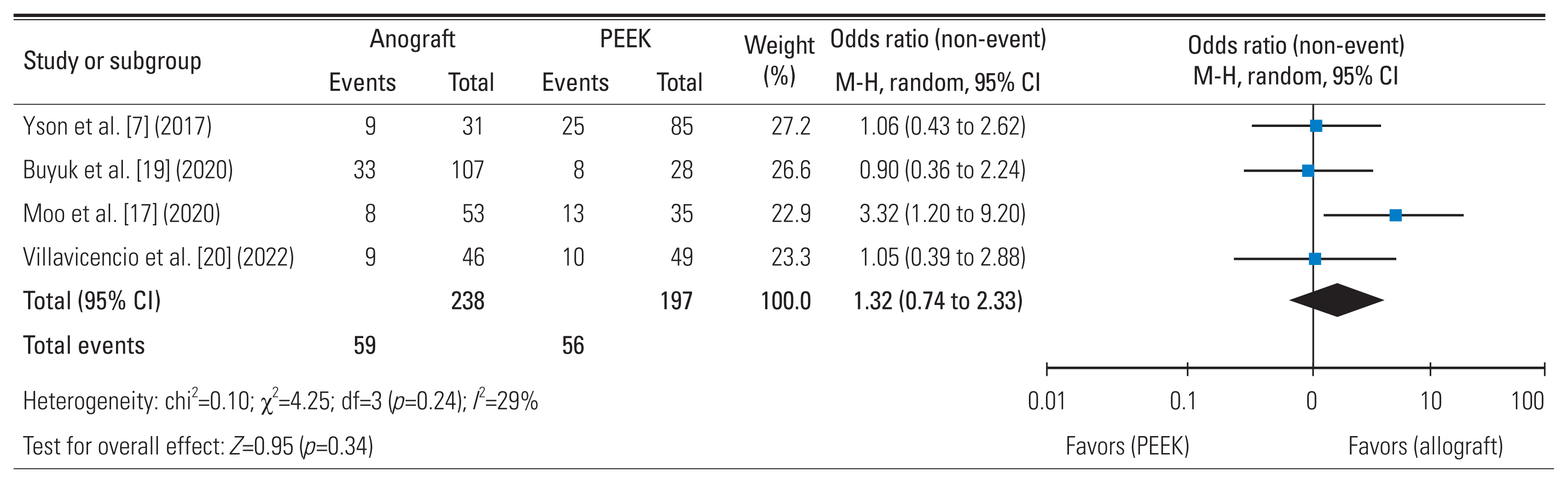
7. Yson SC, Sembrano JN, Santos ER. Comparison of allograft and polyetheretherketone (PEEK) cage subsidence rates in anterior cervical discectomy and fusion (ACDF). J Clin Neurosci 2017 38:118–21.
crossref pmid
8. Yoon ST, Konopka JA, Wang JC, et al. ACDF graft selection by surgeons: survey of AOSpine members. Global Spine J 2017 7:410–6.
crossref pmid pmc pdf
9. Jain A, Marrache M, Harris A, et al. Structural allograft versus PEEK implants in anterior cervical discectomy and fusion: a systematic review. Global Spine J 2020 10:775–83.
crossref pmid pmc pdf
10. Fatima N, Massaad E, Shankar GM, Shin JH. Structural allograft versus polyetheretherketone implants in patients undergoing spinal fusion surgery: a systematic review and meta-analysis. World Neurosurg 2020 136:101–9.
crossref pmid
11. Page MJ, McKenzie JE, Bossuyt PM, et al. The PRISMA 2020 statement: an updated guideline for reporting systematic reviews. BMJ 2021 372:n71.
crossref pmid pmc
12. Stang A. Critical evaluation of the Newcastle-Ottawa scale for the assessment of the quality of nonrandomized studies in meta-analyses. Eur J Epidemiol 2010 25:603–5.
crossref pmid pdf
13. Wan X, Wang W, Liu J, Tong T. Estimating the sample mean and standard deviation from the sample size, median, range and/or interquartile range. BMC Med Res Methodol 2014 14:135.
crossref pmid pmc pdf
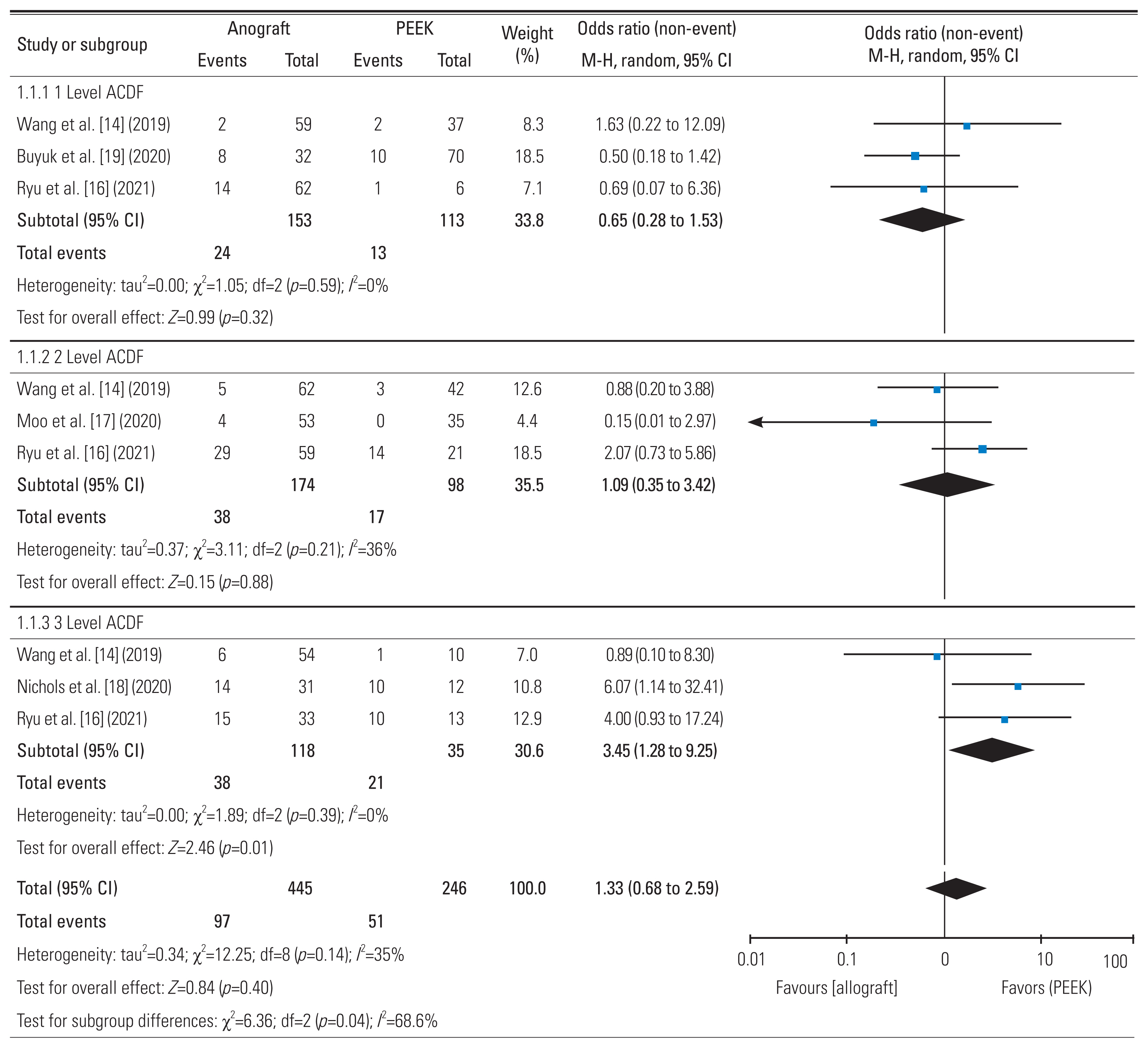
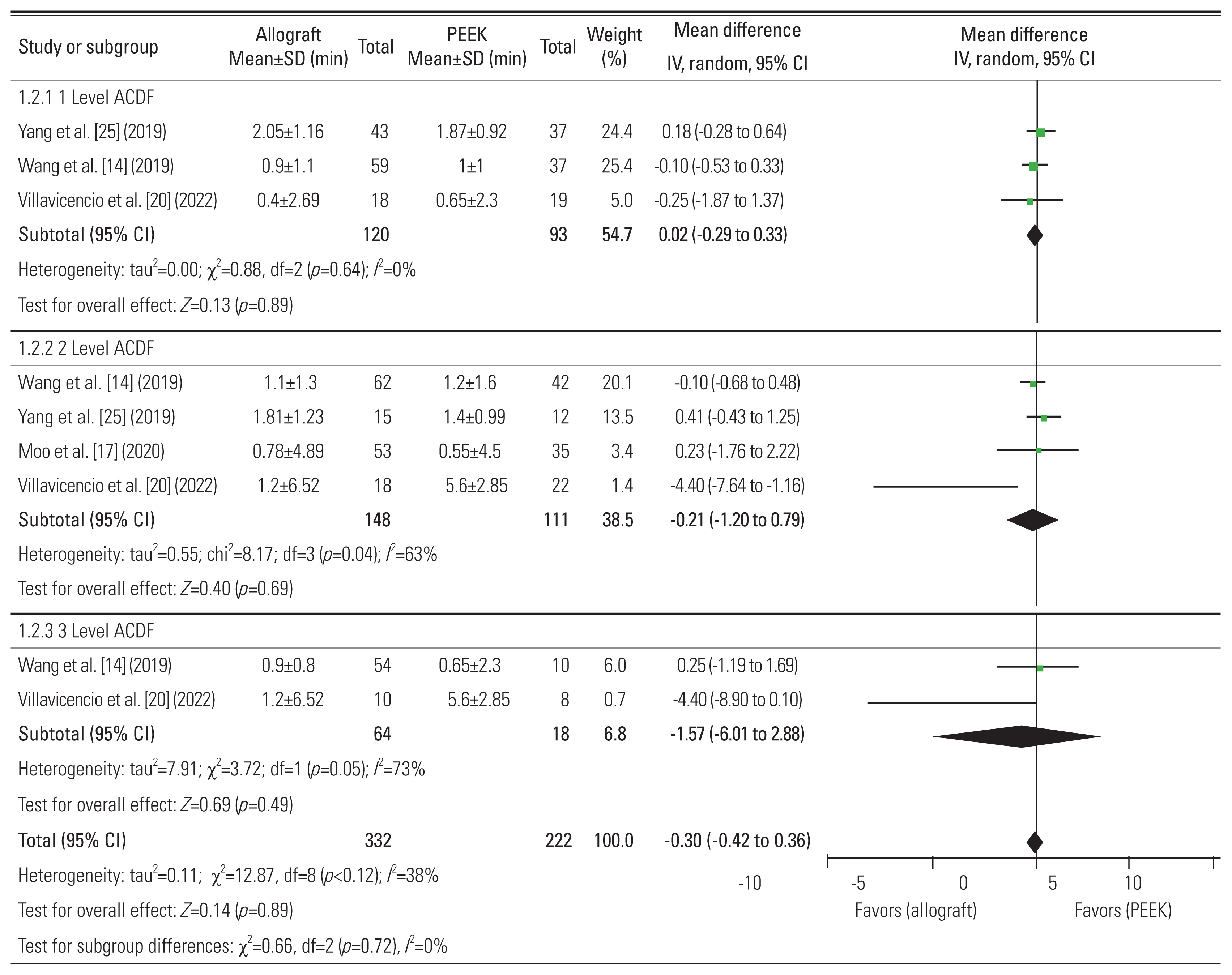
14. Wang M, Chou D, Chang CC, et al. Anterior cervical discectomy and fusion performed using structural allograft or polyetheretherketone: pseudarthrosis and revision surgery rates with minimum 2-year follow-up. J Neurosurg Spine 2019 32:562–9.
crossref
15. Bergin SM, Wang TY, Park C, et al. Pseudarthrosis rate following anterior cervical discectomy with fusion using an allograft cellular bone matrix: a multi-institutional analysis. Neurosurg Focus 2021 50:E6.
crossref
16. Ryu WHA, Richards D, Kerolus MG, et al. Nonunion rates after anterior cervical discectomy and fusion: comparison of polyetheretherketone vs structural allograft implants. Neurosurgery 2021 89:94–101.
pmid
17. Moo IH, Kam CJ, Lai MW, Yeo W, Soh RC. A comparison of contiguous two-level anterior cervical discectomy and fusion using a structural allograft versus a polyetheretherketone (PEEK) cage: the results of a three-year follow-up. BMC Musculoskelet Disord 2020 21:331.
crossref pmid pmc pdf
18. Nichols NM, Jamieson A, Wang M, Chou D, Mummaneni PV, Tan LA. Characterizing the fusion order and level-specific rates of arthrodesis in 3-level anterior cervical discectomy and fusion: a radiographic study. J Clin Neurosci 2020 81:328–33.
crossref pmid
19. Buyuk AF, Onyekwelu I, Gaffney CJ, et al. Symptomatic pseudarthrosis requiring revision surgery after 1- or 2-level ACDF with plating: PEEK versus allograft. J Spine Surg 2020 6:670–80.
crossref pmid pmc
20. Villavicencio AT, Nelson EL, Rajpal S, Beasley K, Burneikiene S. Prospective, randomized, blinded clinical trial comparing PEEK and allograft spacers in patients undergoing anterior cervical discectomy and fusion surgeries. Spine (Phila Pa 1976) 2022 47:1043–54.
crossref pmid
21. Gillis CC, Kaszuba MC, Traynelis VC. Cervical radiographic parameters in 1- and 2-level anterior cervical discectomy and fusion. J Neurosurg Spine 2016 25:421–9.
crossref pmid
22. Langley GB, Sheppeard H. The visual analogue scale: its use in pain measurement. Rheumatol Int 1985 5:145–8.
crossref pmid pdf
23. Vernon H, Mior S. The Neck Disability Index: a study of reliability and validity. J Manipulative Physiol Ther 1991 14:409–15.
pmid
24. Cumpston M, Li T, Page MJ, et al. Updated guidance for trusted systematic reviews: a new edition of the Cochrane Handbook for Systematic Reviews of Interventions. Cochrane Database Syst Rev 2019 10:ED000142.
crossref pmid pmc
25. Yang S, Yu Y, Liu X, et al. Clinical and radiological results comparison of allograft and polyetheretherketone cage for one to two-level anterior cervical discectomy and fusion: a CONSORT-compliant article. Medicine (Baltimore) 2019 98:e17935.
pmid pmc
26. Toth JM, Wang M, Estes BT, Scifert JL, Seim HB, Turner AS. Polyetheretherketone as a biomaterial for spinal applications. Biomaterials 2006 27:324–34.
crossref pmid
27. Kersten RF, van Gaalen SM, de Gast A, Oner FC. Polyetheretherketone (PEEK) cages in cervical applications: a systematic review. Spine J 2015 15:1446–60.
crossref pmid
28. Shimizu T, Fujibayashi S, Yamaguchi S, et al. In vivo experimental study of anterior cervical fusion using bioactive polyetheretherketone in a canine model. PLoS One 2017 12:e0184495.
crossref pmid pmc
29. D’Antonio N, Lambrechts MJ, Heard J, et al. Effect of interbody composition on the development of pseudarthrosis following anterior cervical discectomy and fusion. Asian Spine J 2023 17:518–28.
crossref pmid pmc pdf
30. Delloye C, Cornu O, Druez V, Barbier O. Bone allografts: what they can offer and what they cannot. J Bone Joint Surg Br 2007 89:574–9.
pmid
31. Lee DH, Cho JH, Hwang CJ, et al. What is the fate of pseudarthrosis detected 1 year after anterior cervical discectomy and fusion? Spine (Phila Pa 1976) 2018 43:E23–8.
crossref pmid
32. Olivares-Navarrete R, Gittens RA, Schneider JM, et al. Osteoblasts exhibit a more differentiated phenotype and increased bone morphogenetic protein production on titanium alloy substrates than on poly-ether-ether-ketone. Spine J 2012 12:265–72.
crossref pmid pmc
33. Veeravagu A, Cole T, Jiang B, Ratliff JK. Revision rates and complication incidence in single- and multilevel anterior cervical discectomy and fusion procedures: an administrative database study. Spine J 2014 14:1125–31.
crossref pmid
34. Jiang SD, Jiang LS, Dai LY. Anterior cervical discectomy and fusion versus anterior cervical corpectomy and fusion for multilevel cervical spondylosis: a systematic review. Arch Orthop Trauma Surg 2012 132:155–61.
crossref pmid pdf
35. Bolesta MJ, Rechtine GR, Chrin AM. Three- and four-level anterior cervical discectomy and fusion with plate fixation: a prospective study. Spine (Phila Pa 1976) 2000 25:2040–6.
pmid
36. Steinmetz MP, Warbel A, Whitfield M, Bingaman W. Preliminary experience with the DOC dynamic cervical implant for the treatment of multilevel cervical spondylosis. J Neurosurg 2002 97:330–6.
crossref pmid
37. Tye GW, Graham RS, Broaddus WC, Young HF. Graft subsidence after instrument-assisted anterior cervical fusion. J Neurosurg 2002 97:186–92.
crossref pmid
38. Yue WM, Brodner W, Highland TR. Long-term results after anterior cervical discectomy and fusion with allograft and plating: a 5- to 11-year radiologic and clinical follow-up study. Spine (Phila Pa 1976) 2005 30:2138–44.
pmid
39. van Jonbergen HP, Spruit M, Anderson PG, Pavlov PW. Anterior cervical interbody fusion with a titanium box cage: early radiological assessment of fusion and subsidence. Spine J 2005 5:645–9.
crossref pmid
40. Bartels RH, Donk RD, Feuth T. Subsidence of stand-alone cervical carbon fiber cages. Neurosurgery 2006 58:502–8.
crossref pmid pdf
41. Schmieder K, Wolzik-Grossmann M, Pechlivanis I, Engelhardt M, Scholz M, Harders A. Subsidence of the wing titanium cage after anterior cervical interbody fusion: 2-year follow-up study. J Neurosurg Spine 2006 4:447–53.
crossref pmid
42. Hakalo J, Pezowicz C, Wronski J, Bedzinski R, Kasprowicz M. The process of subsidence after cervical stabilizations by cage alone, cage with plate and plate-cage. A biomechanical comparative study. Neurol Neurochir Pol 2007 41:411–6.
pmid
43. Kurtz SM, Devine JN. PEEK biomaterials in trauma, orthopedic, and spinal implants. Biomaterials 2007 28:4845–69.
crossref pmid pmc
44. Niu CC, Liao JC, Chen WJ, Chen LH. Outcomes of interbody fusion cages used in 1 and 2-levels anterior cervical discectomy and fusion: titanium cages versus polyetheretherketone (PEEK) cages. J Spinal Disord Tech 2010 23:310–6.
pmid
45. Cabraja M, Oezdemir S, Koeppen D, Kroppenstedt S. Anterior cervical discectomy and fusion: comparison of titanium and polyetheretherketone cages. BMC Musculoskelet Disord 2012 13:172.
crossref pmid pmc pdf
46. Das AK, Purohit DK, Gupta A, Kataria R. Comparison of radiological and clinical outcomes between expandable and non-expandable cages following cervical corpectomy: a systematic review and meta-analysis. Asian Spine J 2023 17:567–81.
crossref pmid pmc pdf
47. Karikari IO, Jain D, Owens TR, et al. Impact of subsidence on clinical outcomes and radiographic fusion rates in anterior cervical discectomy and fusion: a systematic review. J Spinal Disord Tech 2014 27:1–10.
pmid
48. Ramakrishna S, Mayer J, Wintermantel E, Leong KW. Biomedical applications of polymer-composite materials: a review. Compos Sci Technol 2001 61:1189–224.
crossref
49. Ma R, Tang T. Current strategies to improve the bioactivity of PEEK. Int J Mol Sci 2014 15:5426–45.
crossref pmid pmc
50. Heary RF, Parvathreddy N, Sampath S, Agarwal N. Elastic modulus in the selection of interbody implants. J Spine Surg 2017 3:163–7.
crossref pmid pmc
51. Carter N, Gianulis EC, Moore MA,Allograft structural interbody spacers compared to PEEK cages in cervical fusion: benchtop and clinical evidence. Barbeck M, Rosenberg N, Rider P, Peric Kacarevic Z, Jung O, editors. Clinical implementation of bone regeneration and maintenance. London: IntechOpen; 2021.Chapter 4: https://doi.org/10.5772/intechopen.88091
crossref
52. Black J, Hastings G. Handbook of biomaterial properties. New York (NY): Springer; 2014.

53. Pennington Z, Mehta VA, Lubelski D, et al. Quality of life and cost implications of pseudarthrosis after anterior cervical discectomy and fusion and its subsequent revision surgery. World Neurosurg 2020 133:e592–9.
crossref pmid
54. Berman D, Oren JH, Bendo J, Spivak J. The effect of smoking on spinal fusion. Int J Spine Surg 2017 11:29.
crossref pmid pmc


ABOUT
ARTICLE CATEGORY

Browse all articles >

BROWSE ARTICLES
EDITORIAL POLICY
FOR CONTRIBUTORS
Editorial Office
Department of Orthopedic Surgery, Asan Medical Center, University of Ulsan College of Medicine
88, Olympic-ro 43-gil, Songpa-gu, Seoul 05505, Korea
Tel: +82-2-3010-3530    Fax: +82-2-3010-8555    E-mail: asianspinejournal@gmail.com                
Korean Society of Spine Surgery
27, Dongguk-ro, Ilsandong-gu, Goyang-si 10326, Korea
Tel: +82-31-966-3413    Fax: +82-2-831-3414    E-mail: office@spine.or.kr                

Copyright © 2024 by Korean Society of Spine Surgery.

Developed in M2PI

Close layer
prev next