Spine Fractures of Patients with Ankylosing Spondylitis and Diffuse Idiopathic Skeletal Hyperostosis: Fracture Severity and Injury-Related Mortality at a Level I Trauma Center
-
Stephen Ryan Chen1,2
 , Maria Amelia Munsch1,2
 , Joseph Chen1,2
 , Brandon Keith Couch1,2
 , Richard Alan Wawrose1,2
 , Anthony Abimbade Oyekan1,2
 , Joshua Adjei1,2
 , William F. Donaldson1,2, Joon Yung Lee1,2, Jeremy DeWitt Shaw1,2
- Received April 18, 2022 Revised July 19, 2022 Accepted August 9, 2022
- ABSTRACT
-
- Study Design
- Retrospective review of prospectively collected cohort.
- Purpose
- To identify differences in treatment and mortality of spine fractures in patients with ankylosing conditions of the spine.
- Overview of Literature
- Ankylosing spondylitis (AS) and diffuse idiopathic skeletal hyperostosis (DISH) are the two most common etiologies of ankylosing spinal disorder (ASD). However, studies on the treatment and outcomes of spine fractures in AS and DISH patients remain few.
- Methods
- Patients presenting with a spine fracture were diagnosed with AS or DISH at a single tertiary care center between 2010 and 2019. We excluded those who lacked cross-sectional imaging or fractures occurring at spinal segments affected by ankylosis, as well as polytraumatized patients. Patient demographics, injury mechanism, fracture level, neurologic status, treatment, and 1-year mortality were recorded. Computed tomography imaging was reviewed by two independent readers and graded according to the indicated AO Spine Injury Classification System. Differences in fracture severity, treatment method, and mortality were examined using Student t-tests, chi-square tests, and two-proportion Z-tests with significance set to p<0.05.
- Results
- We identified 167 patients with spine fracture diagnosed with AS or DISH. Patients with AS had more severe fractures and more commonly had surgery than patients with DISH (p<0.001). Despite these differences, 1-year mortality did not significantly differ between AS and DISH patients (p=0.14).
- Conclusions
- Although patients with AS suffered more severe fractures compared to DISH and more frequently underwent surgery for these injuries, outcomes and 1-year mortality did not differ significantly between the two groups. For patients with ASDs and fractures, outcomes appear similar regardless of treatment modality. Consequently, there may be an opportunity for critical reappraisal of operative indications in ASD and a larger role for nonoperative management in these challenging patients.
- Introduction
- Introduction
Ankylosing spinal disorders (ASDs) are inflammatory processes characterized by pathological spinal ossification affecting approximately 1% of the adult population [1]. These conditions lead to limited range of motion and kyphosis. The resultant kyphosis and loss of flexibility can impact balance and predisposes patients with ASDs to falls [2,3]. Unfortunately, in these individuals, abnormal bony fusion of the spine forms a long lever arm that impairs diffusion of forces within and between vertebrae and can predispose the ankylosed spine to vertebral fracture with even minor trauma [4]. Spinal injuries in patients with ASDs are more likely to be unstable, with a significantly higher rate of spinal cord injury and epidural hematoma [5]. The current dogma suggests that there is increased morbidity and mortality associated with conservative management of spine fractures in patients with ASDs, thus the frequent surgical treatments [4,6,7].Ankylosing spondylitis (AS) and diffuse idiopathic skeletal hyperostosis (DISH) are the two most common etiologies of ASD [8]. While distinct disease processes, AS and DISH present with similar clinical scenarios and fractures [6,9]. AS involves ossification of the entire spine, including the intervertebral disc and the posterior elements. In contrast, ossification in DISH is often more focal and can affect the spine in a non-contiguous manner [10]. Due to the ankylosis of posterior elements seen in AS, it has been suggested that spinal fractures in patients with AS are often more catastrophic than those suffered by their DISH counterparts without significant evidence to support this concept. Moreover, due to the incomplete spinal involvement seen in DISH, spinal fractures may be managed similarly to non-ankylosed spines [11–15]. Despite the unique characteristics of AS and DISH, both ASDs often result in a rigid spine, notable kyphosis, and similar extension injury patterns, which make management clinically challenging [15]. However, few studies have described the fracture characteristics, treatment, and outcomes of patients with AS and DISH fracture. Given the complex pathophysiology and fracture characteristics in patients with ASDs, associated factors and ideal treatment strategies for spinal trauma in patients with AS and DISH must be identified.The present study was aimed to identify differences in fracture severity, operative versus nonoperative management, and 1-year mortality between patients with AS and DISH. It was hypothesized that patients with AS will present with more severe fractures, will undergo more operative intervention, and will have higher 1-year mortality rates than patients with DISH.
- Materials and Methods
- Materials and Methods
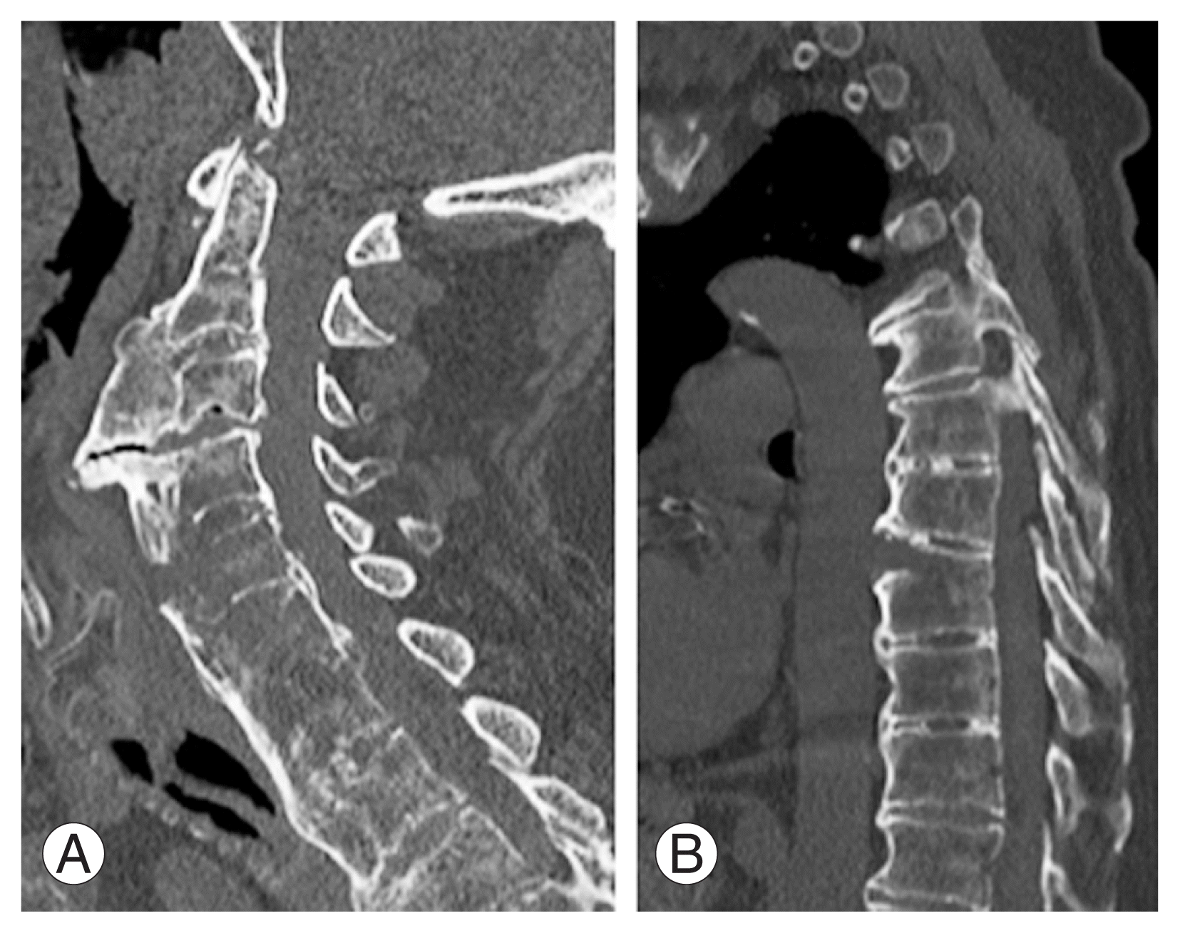
The requirement for informed consent from individual patients was omitted because of the retrospective design of this study. An institutional review board-approved retrospective review of a prospectively collected database was performed on all patients presenting to a single tertiary care center between January 1, 2010, and December 31, 2019, with at least one spinal fracture and a diagnosis of AS or DISH. AS was diagnosed according to the Assessment of SpondyloArthritis international Society classification criteria for axial spondyloarthritis [16,17], while DISH was diagnosed according to the criteria of Resnick and Niwayama [18] as the evidence of ossification of the disc space of AS patients were confirmed by imaging. Imaging was reviewed by three independent reviewers to confirm the diagnosis of AS or DISH fracture (Fig. 1). Exclusion criteria included the absence of appropriate cross-sectional imaging, fractures not graded as A-, B-, or C-type by the AO Spine Injury Classification System (thoracolumbar, subaxial cervical, or upper cervical) [19–21], and fractures occurring outside the spinal segments affected by the ASD. Polytraumatized patients were also excluded.Patient demographics, mechanism of injury, fracture level (cervical, thoracic, thoracolumbar, or lumbar), neurologic status, method of management (operative versus nonoperative), change in long-term mobility status, and mortality (in-hospital, 30-day, 90-day, and 1 year) were collected from the chart. Cervical fractures were defined as injuries extending from the C1 to C7 vertebrae. Thoracic fractures were defined as injuries extending from the T1 to T10 vertebrae. Thoracolumbar fractures were defined as injuries extending from the T11 to L1 vertebrae. Lumbar fractures were defined as injuries extending from the L2 to L5 vertebrae. Treatment modality, specifically operative or nonoperative management and the specifics of surgical treatment or bracing were decided based on clinical condition, fracture characteristics, and neurologic involvement by a fellowship-trained spine surgeon. Operative management specifically entailed spinal fusion with or without decompression. Surgical invasiveness was evaluated according to the spine surgical invasiveness index of Mirza et al. [22].For each patient, computed tomography (CT) imaging of the entire spine was independently reviewed by three independent readers and graded by the previously validated AO Spine Injury Classification System (thoracolumbar, subaxial cervical, or upper cervical) as A-, B-, or C-type fractures [19–21]. Discrepancies in fracture classification were reviewed by the readers and resolved to a consensus grade. Inter-rater reliability was then measured using Cohen’s kappa coefficient [23]. For patients with multiple spinal fractures, the most severe fracture as defined by the AO Spine Injury Classification System was documented and utilized for statistical analysis.Demographic data, AO Spine Injury Classification, treatment modality, and 1-year mortality rates were compared for patients with AS and DISH. Subgroups inclusive of both ASD types were also created based on level of spinal segment injury. The subgroups were compared based on ASD type, AO Spine Injury Classification, treatment modality, and 1-year mortality. Continuous variable comparisons including demographics such as age were performed with Student t-test. Categorical variable differences including demographics or treatment modality proportions were assessed using Pearson’s chi-square test or Fisher’s exact test. Two-proportion Z-test were used to assess differences in mortality rates. A prior power analysis using the 1-year mortality rates found by Meyer [24] and Einsiedel et al. [25] revealed that 92 patients would be required to obtain a power of 80%. Statistical significance was defined as p<0.05.
- Results
- Results
Two-hundred and forty-seven patients with ASD and vertebral fractures were identified (Fig. 2). Eighty patients were eliminated based on the exclusion criteria. Of the remaining 167 patients, 74 patients were diagnosed with AS and 93 patients with DISH. Of them, 123 were male and 44 were female. The mean age at the time of admission was 77.0±12.3 years. The mean age-adjusted Charlson comorbidity index (ACCI) (5.81±2.31 versus 5.04±2.80, p=0.06) and body mass index (29.9±10.4 kg/m2 versus 31.7±12.9 kg/m2, p=0.33) were not significantly different between patients with DISH and patients with AS. Of the study cohort, 135 patients (80.8%) had a low energy mechanism of injury, 162 (97.0%) survived their initial hospital admission, and 121 (72.5%) were followed up, (average follow-up, 8.9 months; range, 0.3–73.6 months). Moreover, 17 patients had multiple non-contiguous fractures; there were 61 cervical, 45 thoracic, 36 thoracolumbar, and 25 lumbar fractures, and 86 A-type, 64 B-type, and 17 C-type fractures. Cohen’s kappa coefficient was 0.62, demonstrating substantial agreement upon fracture classification among raters. Neurologic status classification was not significantly different between AS and DISH patients (p=0.24). Treatment performed on 65 patients was surgical, while on 122 patients was nonsurgical. The mean surgical invasiveness index was greater in patients with AS compared to patients with DISH (14.1±4.8 versus 10.1±4.8, p<0.05). Of the 65 surgically treated patients, 10 (15.4%) had infection at the surgical site. At 1 year after initial presentation, 48 patients (28.7%) died. The mean ACCI was significantly different between living and deceased patients (5.17±2.50 versus 6.21±2.57, p=0.02).Type of ASD significantly correlated with AO Spine Injury Classification (p<0.001) (Table 1). A-type fractures were more frequently seen in DISH fractures than in AS fractures (68.8% versus 29.7%). Conversely, B- and C-type fractures were less frequently seen in DISH fractures than in AS fractures (B-type: 24.7% versus 55.4%; C-type: 6.5% versus 14.9%). Type of ASD also significantly correlated with treatment modality (p<0.001) (Table 2). Patients with DISH fractures were more commonly managed nonoperatively than patients with AS fractures (73.1% versus 45.9%). The type of ASD did not correlate with 1-year mortality (p=0.14) (Table 2). When stratified by spinal segment, AS fractures were more often managed operatively compared to DISH fractures in the thoracic (p<0.05) and thoracolumbar (p<0.05) spine, but there was no significant difference in treatment of cervical (p=0.15) or lumbar (p=0.96) spine fractures (Table 2). At 1-year post-injury, the mortality rate was 28.7% for all patients, 33.3% for patients with DISH, and 23.0% for patients with AS. in the length of stay (p=0.43), in-hospital mortality (p=0.13), 30-day mortality (p=0.48), 90-day mortality (p=0.64), or proportion of patients with reduced long-term mobility (p=0.95) was not significantly different between AS and DISH (Table 2).AO Spine Injury Classification significantly correlated with treatment modality (p<0.001) (Table 3). A-type fractures were more commonly managed nonoperatively (84.9% versus 15.1%), while B- and C-type fractures, operatively (B-type: 39.1% versus 60.9%; C-type: 23.5% versus 76.5%). This trend was consistent among patients with DISH (p<0.001) (Table 3) and patients with AS (p<0.05) (Table 3).AO Spine Injury Classification did not correlate with 1-year mortality (p=0.72) (Table 3). Similarly, there was no correlation between AO Spine Injury Classification and one-year mortality for patients with DISH (p=0.21) (Table 3) and patients with AS (p=0.50) (Table 3). In proportional assessment of all ASD types, 1-year mortality was greatest among A-type (31.4%) fractures, followed by B-type (26.6%) and C-type fractures. Patients treated nonoperatively had a similar 1-year mortality compared to patients treated operatively (31.4% versus 24.6%, p=0.35) (Table 4). Operative as compared to nonoperative management had no significant differences in length of stay, in-hospital mortality, 30-day mortality, 90-day mortality, or loss of long-term mobility (all p>0.13) (Table 4).
- Discussion
- Discussion
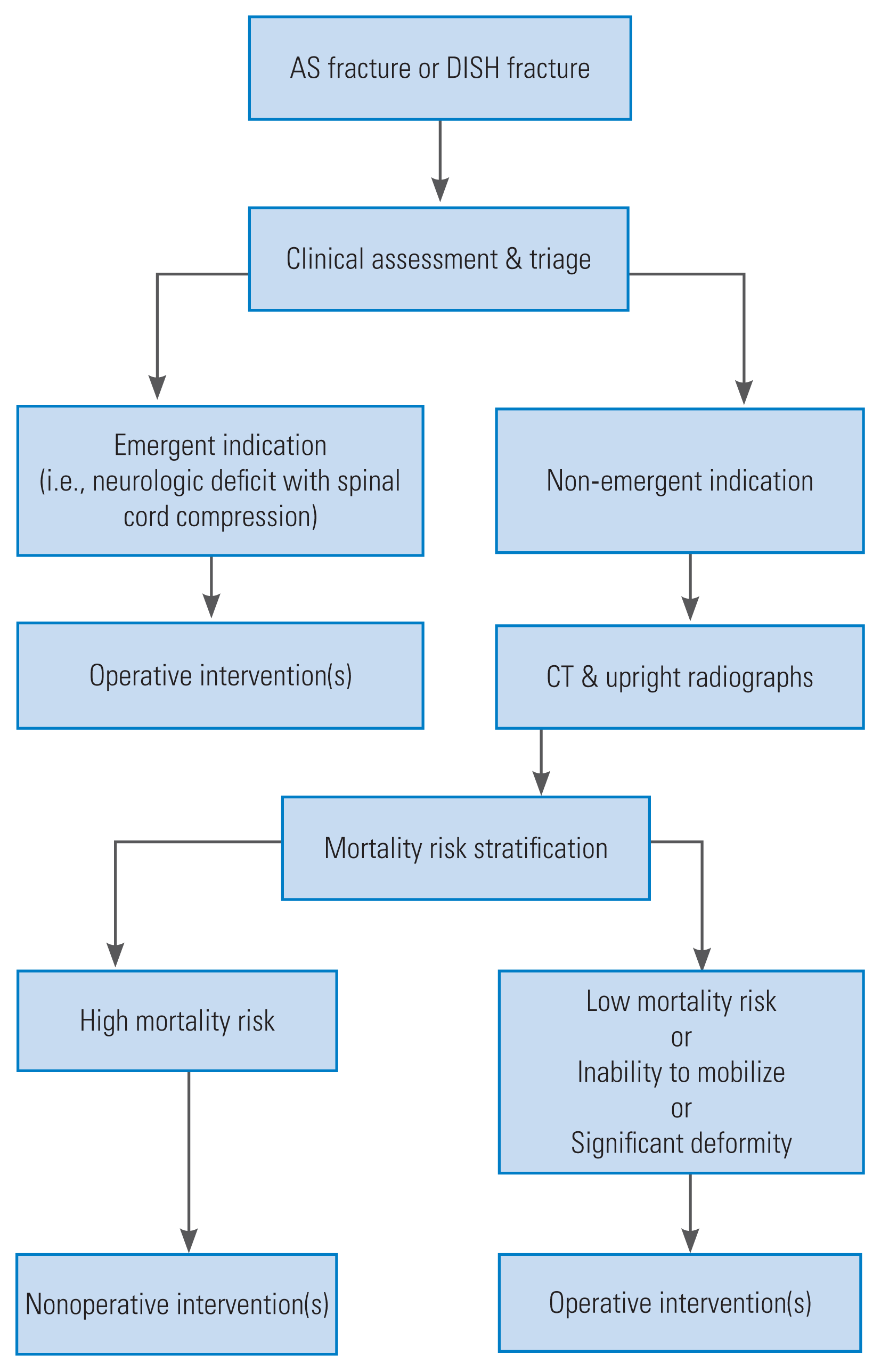
The distinct pathophysiology between AS and DISH results in unique injury characteristics in patients with spinal trauma. The purpose of this study was to identify differences between types of ASD with respect to fracture severity, treatment modality, and 1-year mortality rates. While patients with AS had more severe fractures and underwent operative intervention more frequently than patients with DISH, the type of ASD did not affect 1-year mortality rates. As expected, more severe fractures were more likely to undergo operative intervention. However, neither fracture severity nor treatment modality predicted 1-year mortality rates.Overall, this patient cohort and their injury patterns are consistent with those reported in previously published literature. The average age of the patients was 77 years, which is slightly older than the 45–76 years reported by Rustagi et al. [26] in their meta-analysis. However, most of the studies included in the meta-analysis focused solely on patients with AS, which have a younger average age at time of fracture than patients with DISH [4,6]. The larger number of patients with DISH included in this sample may account for the older average patient age; 73.7% of the patient cohort were males, which agrees with the fact that ASDs predominantly affect men [4,6,7]; 80.8% of the patients sustained a low-energy mechanism of injury, which is slightly higher than the 64%–78% previously reported [4,7,13]. However, the exclusion of polytraumatized patients from this patient cohort likely eliminated more patients with high-energy mechanisms of injury. Approximately 10% of patients suffered fractures in non-contiguous vertebrae, which is also similar to the previously reported data [6,27]. These findings suggest that this cohort was broadly representative of patients with ASDs that experienced isolated spinal injury.In general, fractures which are classified as more severe by the AO Spine Injury Classification system are more likely to warrant operative intervention and portend worse long-term outcomes [28,29]. Although not significant, the data demonstrate a paradoxical trend in mortality as A-type and B-type fractures had greater mortality than C-type fractures. However, this trend may be explained by ACCI for the deceased patients, with ACCI of 6.85, 5.47, and 5.00 for A-type, B-type, and C-type fractures, respectively. Additionally, this may have led to inadvertent selection bias as a portion of fracture patients are treated nonoperatively based on increased medical complexity and consequent risk of perioperative morbidity and mortality rather than based on fracture morphology alone.In this cohort, isolated spinal fractures are more likely to undergo operative intervention in the setting of AS than in patients with DISH [13,30]. This may have been due to the prevalence of posterior element injury and the distribution of fractures which where AS fractures were more severe in nature than DISH fractures. Stratification by spinal segment demonstrates an interesting trend for DISH fractures. While cervical fractures underwent a similar rate of nonoperative and operative management, fractures at all other levels were more likely to be treated nonoperatively. This differential treatment is likely multifactorial, however, may be due in part to fixed kyphosis of the cervical spine, which complicates immobilization by bracing and makes operative intervention a more attractive option [9]. The sternal-rib complex of the thoracic spine imparts additional stability to those vertebrae which often makes thoracic spine fractures good candidates for nonoperative management [31]. Lastly, as the spinal cord generally terminates in the upper lumbar region there is lower risk of neurologic injury in for injuries in the lumbar spine.Despite the differences in treatment between AS and DISH, the data seems to suggest that one-year mortality is not influenced by type of ASD or treatment modality. In fact, patients with DISH demonstrated similar mortality regardless of treatment. Although not significant, the data demonstrates improved mortality for patients with DISH that are treated nonoperatively. The mortality benefit of nonoperative management in patients with DISH may be due in part due to the relatively high rate of surgical site infection in this cohort. The paradoxical mortality benefit of nonoperative management encourages careful consideration of indications for surgery as every procedure has risk of iatrogenic injury, implant failure, and infection. For patients with AS, although not significant, operative treatment showed a trend toward improved mortality over nonoperative treatment.At 1-year post-injury, there was a significant difference between living and deceased patients regarding the burden of medical comorbidities as measured by the ACCI at the time of injury. The ACCI may provide prognostic information that helps to guide treatment decisions. This further suggests that perioperative risk stratification is imperative for good surgical outcomes in this high-risk patient population. A suggested management pathway prioritizes nonoperative management for all ASD fractures unless specifically indicated by progressive deficit or significant fracture associated deformity (Fig. 3). While a substantial number of patients were treated with surgery, bracing with pain control and early mobilization was the standard for this cohort unless a clear operative indication existed.This study has some limitations, due largely to its retrospective nature. The selection of patients for surgical management may have obscured existing associations between treatment modality underlying patient medical comorbidity. For example, patients with baseline higher mortality risk, are more likely to be managed nonoperatively. Additionally, patients with DISH were older than the patients with AS. Age is a known predictor of mortality in fractures with ASD [6]. Regardless of age differences in this cohort, the burden of medical comorbidities as measured by the ACCI did not significantly differ between patients with DISH and AS, strengthening comparisons between the two groups. Despite these limitations, this patient cohort of 167 total patients is larger than most other studies on ASD fractures and power analysis suggests adequate powering to detect differences between the two cohorts. In addition, the exclusion of polytraumatized patients likely strengthens this study as it limits confounding medical and surgical complexities from analyses.
- Conclusions
- Conclusions
Although patients with AS suffered more severe fractures when compared to DISH and more frequently underwent surgery for these injuries, outcomes and one-year mortality were not different between the two groups. While operative fixation is considered the dogmatic standard for fractures in the ankylosed spine, outcomes at 1 year suggest more clinical equipoise between operative or nonoperative management of fractures in patients with ASDs than has previously been reported. Consequently, there may be an opportunity for critical reappraisal of operative indications in ASD and a larger role for nonoperative management in these challenging patients. A trial of bracing, pain control, and early mobilization is suggested for all patients with ASD fracture unless in case of emergent surgical indications or an eventual failure of nonoperative management. Future prospective studies should identify a treatment pathway to establish greater insight into the ideal treatment strategy for patients with ASDs.
- Notes
- Notes
-
Conflict of Interest No potential conflict of interest relevant to this article was reported.
Author Contributions Study conception: JDS; manuscript preparation: SRC, MAM; development of methodology: SRC; data collection: JC, BKC, RAW, JA; data curation: JC, BKC; computation: RAW; data analysis: SRC, AAO; manuscript writing: SRC; critical review: MAM, AAO, JYL, JDS; project administration: WFD, JYL, JDS; and supervision: WFD, JYL, JDS.
Fig. 1

Fig. 2

Fig. 3

Table 1
Values are presented as number, mean±standard deviation, or number (%). Eligible patients are stratified according to their type of ankylosing spinal disorder. p-values are from the Student t-test for quantitative variables and from the Pearson’s chi-square test or Fisher’s exact for categorical variables comparing AS to DISH.
Table 2
| Characteristic | All study participants | DISH | AS | p-value |
|---|---|---|---|---|
| No. of patients | 167 | 93 | 74 | |
| Fracture region | ||||
| Cervical (C1–C7) | 61 | 32 | 29 | 0.15 |
| Nonoperative | 29 (47.5) | 18 (56.3) | 11 (37.9) | |
| Operative | 32 (52.5) | 14 (43.7) | 18 (62.1) | |
| Thoracic (T1–T10) | 45 | 22 | 23 | <0.05 |
| Nonoperative | 31 (68.9) | 19 (86.4) | 12 (52.2) | |
| Operative | 14 (31.1) | 3 (13.6) | 11 (47.8) | |
| Thoracolumbar (T11–L1) | 36 | 22 | 14 | <0.05 |
| Nonoperative | 20 (55.6) | 16 (72.7) | 4 (28.6) | |
| Operative | 16 (44.4) | 6 (27.3) | 10 (71.4) | |
| Lumbar (L2–L5) | 25 | 17 | 8 | 0.96 |
| Nonoperative | 22 (88.0) | 15 (88.2) | 7 (87.5) | |
| Operative | 3 (12.0) | 2 (11.8) | 1 (12.5) | |
| Treatment | <0.001 | |||
| Nonoperative | 102 (61.1) | 68 (73.1) | 34 (45.9) | |
| Operative | 65 (38.9) | 25 (26.9) | 40 (54.1) | |
| Surgical site infection | 10 (15.4) | 3 (12.0) | 7 (17.5) | 0.54 |
| Surgical invasiveness index | 12.6±5.2 | 10.1±4.8 | 14.1±4.8 | <0.05 |
| Length of stay (day)a) | 7.6±6.2 | 7.3±6.4 | 8.0±5.8 | 0.43 |
| Loss of mobilitya) | 49 (30.2) | 28 (17.3) | 21 (13.0) | 0.95 |
| In-hospital mortality | 5 (3.0) | 1 (1.1) | 4 (5.4) | 0.13 |
| 30-Day mortality | 13 (7.8) | 6 (6.5) | 7 (9.5) | 0.48 |
| 90-Day mortality | 25 (15) | 15 (16.5) | 10 (13.5) | 0.64 |
| 1-Year mortality | 48 (28.7) | 31 (33.3) | 17 (23.0) | 0.14 |
Values are presented as number, mean±standard deviation, or number (%). Eligible patients are stratified according to their type of ankylosing spinal disorder. Categorized by region of fracture and therapeutic intervention. Surgical invasiveness index calculated according to Mirza et al. [22]. Loss of mobility describes long-term need for assistive device, wheelchair, or bedridden status which was not present at baseline. Below are mortality rates stratified by intervention. p-values are from two proportion Z-test for mortality, the Student t-test for quantitative variables, and Pearson’s chi-square test or Fisher’s exact for categorical variables comparing AS to DISH.
Table 3
Values are presented as number, number (%), or % (mortality/number). Eligible patients are stratified according to their AO classification fracture pattern and type of ankylosing spinal disorder. Mortality rates are presented as proportions. p-values are from the Pearson’s chi-square test or Fisher’s exact for categorical variables and two proportion Z-test for mortality. Comparisons are between fracture patterns.
Table 4
| Characteristic | All study participants | Nonoperative | Operative | p-value |
|---|---|---|---|---|
| No. of patients | 167 | 102 | 65 | |
| 1-Year mortality rate | 28.7 (48/167) | 31.4 (32/102) | 24.6 (16/65) | 0.35 |
| AS | 22.7 (17/74) | 32.4 (11/34) | 15.0 (6/40) | 0.41 |
| DISH | 33.3 (31/93) | 30.9 (21/68) | 40.0 (10/25) | 0.08 |
| In-hospital mortality | 3.0 (5/167) | 3.9 (4/102) | 1.5 (1/65) | 0.33 |
| 30-Day mortality | 7.8 (13/167) | 8.8 (9/102) | 6.2 (4/65) | 0.51 |
| 90-Day mortality | 15 (25/167) | 15.7 (16/102) | 13.8 (9/65) | 0.74 |
| Length of stay (day)a) | 7.6±6.2 | 7.9±6.3 | 7.1±6.3 | 0.45 |
| Loss of mobilitya) | 30.2 (49/162) | 32.3 (32/99) | 27.0 (17/63) | 0.47 |
Values are presented as number, % (affected number/number), or mean±standard deviation. Eligible patients are stratified according to their operative or nonoperative management and type of ankylosing spinal disorder. Mortality rates are presented as proportions. Loss of mobility describes long-term need for assistive device, wheelchair, or bedridden status which was not present at baseline. p-values are from two proportion Z-test for mortality, chi-square for loss of mobility, and Student t-test for length of stay comparing operative vs. nonoperative management.
- REFERENCES
- REFERENCES
References
1. Taurog JD, Chhabra A, Colbert RA. Ankylosing spondylitis and axial spondyloarthritis. N Engl J Med 2016;374:2563–74.
[Article] [PubMed]2. Dursun N, Sarkaya S, Ozdolap S, et al. Risk of falls in patients with ankylosing spondylitis. J Clin Rheumatol 2015;21:76–80.
[Article] [PubMed]3. Vergara ME, O’Shea FD, Inman RD, Gage WH. Postural control is altered in patients with ankylosing spondylitis. Clin Biomech (Bristol, Avon) 2012;27:334–40.
[Article] [PubMed]4. Westerveld LA, Verlaan JJ, Oner FC. Spinal fractures in patients with ankylosing spinal disorders: a systematic review of the literature on treatment, neurological status and complications. Eur Spine J 2009;18:145–56.
[Article] [PubMed] [PMC]5. Alaranta H, Luoto S, Konttinen YT. Traumatic spinal cord injury as a complication to ankylosing spondylitis: an extended report. Clin Exp Rheumatol 2002;20:66–8.
[PubMed]6. Caron T, Bransford R, Nguyen Q, Agel J, Chapman J, Bellabarba C. Spine fractures in patients with ankylosing spinal disorders. Spine (Phila Pa 1976) 2010;35:E458–64.
[Article] [PubMed]7. Lu ML, Tsai TT, Lai PL, et al. A retrospective study of treating thoracolumbar spine fractures in ankylosing spondylitis. Eur J Orthop Surg Traumatol 2014;24(Suppl 1): S117–23.
[Article] [PubMed]8. Hartmann S, Tschugg A, Wipplinger C, Thome C. Analysis of the literature on cervical spine fractures in ankylosing spinal disorders. Global Spine J 2017;7:469–81.
[Article] [PubMed] [PMC]9. Reinhold M, Knop C, Kneitz C, Disch A. Spine fractures in ankylosing diseases: recommendations of the spine section of the German Society for Orthopaedics and Trauma (DGOU). Global Spine J 2018;8(2 Suppl): 56S–68S.
[Article] [PubMed] [PMC]10. Olivieri I, D’Angelo S, Palazzi C, Padula A, Mader R, Khan MA. Diffuse idiopathic skeletal hyperostosis: differentiation from ankylosing spondylitis. Curr Rheumatol Rep 2009;11:321–8.
[Article] [PubMed]11. Okada E, Tsuji T, Shimizu K, et al. CT-based morphological analysis of spinal fractures in patients with diffuse idiopathic skeletal hyperostosis. J Orthop Sci 2017;22:3–9.
[Article] [PubMed]12. Schoenfeld AJ, Harris MB, McGuire KJ, Warholic N, Wood KB, Bono CM. Mortality in elderly patients with hyperostotic disease of the cervical spine after fracture: an age- and sex-matched study. Spine J 2011;11:257–64.
[Article] [PubMed]13. Shah NG, Keraliya A, Harris MB, Bono CM, Khurana B. Spinal trauma in DISH and AS: is MRI essential following the detection of vertebral fractures on CT? Spine J 2021;21:618–26.
[Article] [PubMed]14. Tan T, Huang MS, Hunn MK, Tee J. Patients with ankylosing spondylitis suffering from AO type B3 traumatic thoracolumbar fractures are associated with increased frailty and morbidity when compared with patients with diffuse idiopathic skeletal hyperostosis. J Spine Surg 2019;5:425–32.
[Article] [PubMed] [PMC]15. Whang PG, Goldberg G, Lawrence JP, et al. The management of spinal injuries in patients with ankylosing spondylitis or diffuse idiopathic skeletal hyperostosis: a comparison of treatment methods and clinical outcomes. J Spinal Disord Tech 2009;22:77–85.
[PubMed]16. Correction: the development of assessment of spondyloArthritis international society classification criteria for axial spondyloarthritis (part II): validation and final selection. Ann Rheum Dis 2019;78:e59.
[Article]17. Bakland G, Alsing R, Singh K, Nossent JC. Assessment of spondyloarthritis international society criteria for axial spondyloarthritis in chronic back pain patients with a high prevalence of HLA-B27. Arthritis Care Res (Hoboken) 2013;65:448–53.
[Article] [PubMed]18. Resnick D, Niwayama G. Radiographic and pathologic features of spinal involvement in diffuse idiopathic skeletal hyperostosis (DISH). Radiology 1976;119:559–68.
[Article] [PubMed]19. Vaccaro AR, Koerner JD, Radcliff KE, et al. AOSpine subaxial cervical spine injury classification system. Eur Spine J 2016;25:2173–84.
[Article] [PubMed]20. Vaccaro AR, Oner C, Kepler CK, et al. AOSpine thoracolumbar spine injury classification system: fracture description, neurological status, and key modifiers. Spine (Phila Pa 1976) 2013;38:2028–37.
[PubMed]21. Kepler CK, Vaccaro AR, Koerner JD, et al. Reliability analysis of the AOSpine thoracolumbar spine injury classification system by a worldwide group of naive spinal surgeons. Eur Spine J 2016;25:1082–6.
[Article] [PubMed]22. Mirza SK, Deyo RA, Heagerty PJ, et al. Development of an index to characterize the “invasiveness” of spine surgery: validation by comparison to blood loss and operative time. Spine (Phila Pa 1976) 2008;33:2651–62.
[PubMed]23. Cohen J. A coefficient of agreement for nominal scales. Educ Psychol Meas 1960;20:37–46.
[Article]24. Meyer PR Jr.. Diffuse idiopathic skeletal hyperostosis in the cervical spine. Clin Orthop Relat Res 1999;(359): 49–57.
[Article]25. Einsiedel T, Schmelz A, Arand M, et al. Injuries of the cervical spine in patients with ankylosing spondylitis: experience at two trauma centers. J Neurosurg Spine 2006;5:33–45.
[Article] [PubMed]26. Rustagi T, Drazin D, Oner C, et al. Fractures in spinal ankylosing disorders: a narrative review of disease and injury types, treatment techniques, and outcomes. J Orthop Trauma 2017;31(Suppl 4): S57–74.
[Article] [PubMed]27. Osgood CP, Abbasy M, Mathews T. Multiple spine fractures in ankylosing spondylitis. J Trauma 1975;15:163–6.
[Article] [PubMed]28. Aarabi B, Oner C, Vaccaro AR, Schroeder GD, Akhtar-Danesh N. Application of AOSpine subaxial cervical spine injury classification in simple and complex cases. J Orthop Trauma 2017;31(Suppl 4): S24–32.
[Article] [PubMed]29. Vaccaro AR, Schroeder GD, Kepler CK, et al. The surgical algorithm for the AOSpine thoracolumbar spine injury classification system. Eur Spine J 2016;25:1087–94.
[Article] [PubMed]30. Jacobs WB, Fehlings MG. Ankylosing spondylitis and spinal cord injury: origin, incidence, management, and avoidance. Neurosurg Focus 2008;24:E12.
[Article]31. Shen FH, Samartzis D. Successful nonoperative treatment of a three-column thoracic fracture in a patient with ankylosing spondylitis: existence and clinical significance of the fourth column of the spine. Spine (Phila Pa 1976) 2007;32:E423–7.
[PubMed]
