Introduction
The surgical correction of adolescent idiopathic scoliosis (AIS) aims to fuse the structural curves with the least number of fusion levels possible while maintaining a balanced body. However, it had been recognized that in some cases, the unfused compensatory curves above or below the main structural curve are unable to compensate after surgical correction, and ŌĆ£letting goŌĆØ of the initial curve correction was required to achieve a balanced shoulder and pelvis [1]. The flexibility of the proximal thoracic (PT) segments plays an important role in determining whether the surgical correction of the main thoracic (MT) curve will lead to neck and shoulder imbalance [2-4]. An imbalanced neck or shoulder after surgery may result in cosmetic dissatisfaction among patients [5]. As the selection of fusion levels determines the alignment and compensation of the unfused segments, the flexibility of the PT segment may govern the selection of the upper instrumented vertebra during corrective surgery.
Although some authors have stressed the importance of recognizing the rigidity and structurality of the PT curve prior to surgical correction [6], the results of these studies are based on the evaluation of conventional whole spine and supine side-bending radiographs. In the literature, several methods of radiographically assessing the flexibility of the main scoliotic curve using supine bending films, axial suspension films, push-prone films, push-traction films, fulcrum bending films, and traction films have been reported; however, these studies have not investigated PT flexibility [7-14].
Kirk et al. [15] investigated the flexibility of the PT curve by comparing the supine traction radiograph with the conventional supine side-bending radiograph. They found that greater flexibility of the curve was visualized with the supine traction radiograph in comparison to the supine side-bending radiograph. Cervical supine sidebending (CSSB) radiographs are films that combine supervised bending of the cervical spine and bending of the whole spine in the supine position [16]. It is postulated that the CSSB radiographs provide a better assessment of the flexibility of the PT curve in comparison to the conventional supine side-bending radiographs or even the supine traction radiographs. Therefore, the present study analyzed and compared the ability of CSSB and cervical supine traction (CST) radiographs to determine the flexibility and correctability of the PT curve in patients with Lenke 1 and 2 AIS.
Materials and Methods
This prospective clinicalŌĆōradiological study was conducted between December 2015 and June 2016. Ethical approval from University Malaya Medical Centre review board was obtained (Ethical approval no., MECID No. 20159-1630). Inclusion criteria for this study were patients diagnosed with AIS with Lenke 1 or 2 curve who were candidates for posterior spinal fusion. Patients with nonidiopathic scoliosis and revision cases were excluded.
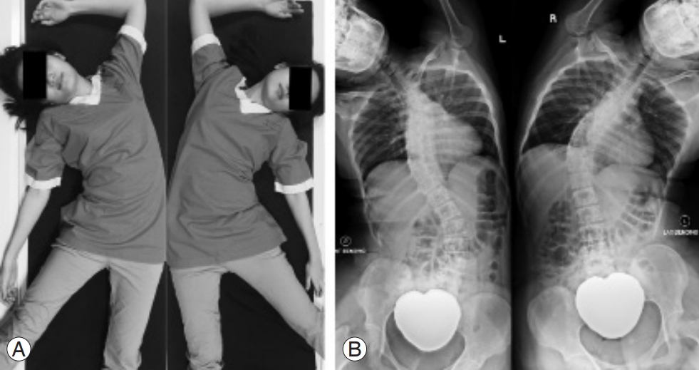
All patients underwent routine preoperative radiography, including standing anteroposterior whole spine, standing lateral whole spine, and supine right and left side-bending radiographs. To analyze the flexibility of the PT segments, two physician-supervised radiographs were obtained: (1) CSSB radiograph: a radiograph taken by bending the cervical spine maximally, performed together with bending the whole spine in the supine position. Right and left CSSB radiographs were obtained by bending the cervical spine and the PT segments to the right and left passively while maintaining the head and neck in neutral rotation (Fig 1). (2) CST radiograph: a PT radiograph taken during traction of the cervical and PT spines with a halter traction device. The traction cranially was counterattracted caudally (Fig. 2).
Patient demographic and radiological parameters included age, gender, weight, height, body mass index, PT angle, MT angle, CSSB PT angle, CST PT angle, and postoperative PT angle. All measurements were digitally taken using a software (Centricity PACS, ver. 5.0; GE Healthcare, Milwaukee, WI, USA). These parameters were measured in accordance with the ŌĆ£Spinal Deformity Study Group radiographic measurement manualŌĆØ [17]. For the PT angle measurement, the uppermost vertebra of the MT curve was used as the lowest vertebra of the PT curve, and T1 or the most tilted vertebra below T1 was used as the uppermost vertebra of the PT curve.
Definitions and calculations of parameters were as follows: (1) CSSB flexibility (CSSBF)=(PT angle.CSSB PT angle)/(PT angle)├Ś100; (2) CST flexibility (CSTF)=(PT angle.CST PT angle)/(PT angle)├Ś100; (3) correction rate (CR)=(PT angle.postoperative PT angle)/(PT angle)├Ś100; (4) CSSB correction index (CSSBCI)=CR/CSSBF; and (5) CST correction index (CSTCI)=CR/CSTF.
Explanations of the calculated parameters were as follows. (1) Flexibility: as the value of this parameter increases, it indicates increased flexibility of the PT scoliotic curve. (2) CR: the amount of PT curve correction achieved after surgery. (3) Correction index (CI): a ratio/index to denote how accurately the flexibility assessment reflects the final surgical correction of the PT scoliotic curve. A value of 1 indicates that the preoperative flexibility assessment is the same as the postoperative CR. A value higher than 1 indicates that the surgical correction is more than the assessed flexibility. A value of less than 1 indicates that the surgical correction is less than the assessed flexibility.
1. Sample size analysis
By using one formula for mean of the sample size estimation, CSSB and CST angles were used as outcome variables in this study. Based on the previous study by Kirk et al. [15], the mean for supine side-bending and supine traction Cobb angles of the PT curve were 30.1┬░ and 24.4┬░, respectively. The largest sample size was selected among those angles. Thus, the effect size was obtained as 0.47 at 80% power of the study, indicating that a minimum sample size of 29 subjects was required to detect the differences in Cobb angles. The calculation was performed using G*Power software (ver. 3.1.9.2; Heinrich-Heine-Universitat Dusseldorf, Dusseldorf, Germany; http://www.gpower.hhu.de/) [18].
2. Statistical analysis
Data was entered and analyzed using IBM SPSS ver. 23.0 statistical software (IBM Corp., Armonk, NY, USA). Descriptive analysis was performed using One-way analysis of variance for quantitative variables and the Žć2 test for categorical variables. The strength of correlation was assessed between CSSBF versus CR, CSTF versus CR, CSSB PT angle versus postoperative PT angle, and CST PT angle versus postoperative PT angle for each subgroup and the whole group using PearsonŌĆÖs correlation coefficient. A paired t-test was used to determine the differences between CSSBF and CSTF, CSSBCI and CSTCI, and CSSB PT angle and CST PT angle within each group. An ╬▒ level of 0.05 indicated statistical significance.
Results
This study recruited 30 patients (26 females, four males) with a mean age of 15.3┬▒3.3 years. The weight, height, body mass index, PT angle, MT angle, postoperative PT angle, CR, and upper instrumented vertebrae (UIV) are illustrated in Table 1.
The preoperative and postoperative measurements are illustrated in Table 2. Both mean CSSB PT (16.6┬░┬▒10.4┬░) and CST PT (23.7┬░┬▒10.7┬░) angles were less than the preoperative standing PT angle (29.4┬░┬▒11.2┬░) and more than the postoperative PT angle (16.1┬░┬▒7.5┬░). Most of the UIV were T3. The UIV was generally higher (T2) when there was less flexibility and lower (T4 or T5) when there was greater flexibility.
There were 15 patients (50%) with a PT angle <15┬░ with CSSB in comparison to five patients (16.7%) with CST. There were seven patients (23.3%) with a PT angle of 15┬░ŌĆō25┬░ with CSSB in comparison to 12 patients (40.0%) with CST. There were eight patients (26.7%) with a PT angle >15┬░ with CSSB in comparison to 13 patients (43.3%) with CST. There were significant differences between PT angles for CSSB and CST (p<0.01).
The Cobb angle, flexibility, and CI comparisons between the CSSB and CST radiographs are illustrated in Table 3. The CSSB PT angle (16.6┬░┬▒10.4┬░) was significantly (p<0.05) smaller than the CST PT angle (23.7┬░┬▒10.7┬░). The CSSBF (44.2%┬▒19.7%) was significantly (p<0.05) greater than the CSTF (19.5%┬▒18.1%). The CSSBCI (1.2┬▒0.9) was significantly (p<0.05) closer to the value 1 in comparison to the CSTCI (4.4┬▒5.3). There was no significant difference (p=0.72) between the CSSB PT angle (16.6┬░┬▒10.4┬░) and the postoperative PT angle (16.1┬░┬▒7.5┬░), but the CST PT angle (23.7┬░┬▒10.7┬░) was significantly (p<0.05) larger than the postoperative PT angle (16.1┬░┬▒7.5┬░).
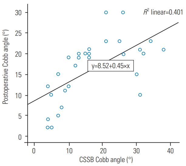
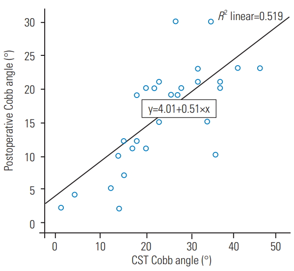
The correlation between the postoperative PT angle and the CSSB PT angle is illustrated in Fig. 3. The correlation between the postoperative PT angle and the CST PT angle is illustrated in Fig. 4. Both the CSSB and CST PT angles are significantly correlated with the postoperative PT angles (p<0.05).
Discussion
Flexibility assessment of a scoliotic curve in a patient with AIS prior to corrective surgery is important. Several methods of radiographically assessing the flexibility of the main scoliotic curve have been reported [7-14]. Some authors report better assessment of the flexibility of the scoliotic curve with traction under general anesthesia [19,20]. However, these reports have only investigated the flexibility of the main scoliotic curve, and none have assessed the flexibility of the PT curve.
The flexibility of the PT curve is an important factor in determining neck and shoulder balance following correction of the MT curve. Neck and shoulder balance following surgery has been studied and reported by many authors. This imbalance can be divided into lateral or medial shoulder imbalance [2-4,21-26]. Coracoid height difference, clavicle rib-intersection difference, clavicular angle, and radiological shoulder height contribute to the lateral shoulder imbalance [3,21-25]. T1 tilt, cervical axis, trapezoidal prominence, and neck tilt contribute to the medial shoulder imbalance [2,4,26]. The lateral shoulder imbalance can usually be compensated by posture adjustments; however, the medial shoulder imbalance does not respond to posture adjustment and should be resolved during corrective surgery.
To avoid neck and shoulder imbalance, several authors have recommended strategies in their selection of the upper instrumented vertebra. Rose and Lenke [27] recommended fusion to T2 if the left shoulder was higher preoperatively, fusion to T2 or T3 if the shoulder was at level, and fusion to T3 if the right shoulder was higher. Suk et al. [28] recommended that if the PT curve was more than 25┬░ with a level or elevated left shoulder, it should be treated as a structural curve with fusion up to T1. Elfilky et al. [29] recommended fusion into the PT curve only when it was more than 45┬░, and a non-fusion strategy was appropriate if the curve was less than 45┬░. Matsumoto et al. [30] found that the mean postoperative clavicular angle was satisfactory even when a short fusion (UIV at one level below the end vertebra) was used for patients with Lenke 1 AIS.
Kirk et al. [15] compared the ability of supine traction radiography and supine side-bending radiography to assess the flexibility of the PT curve in 15 patients with King V/Lenke 2 AIS. They found that a supine traction radiograph demonstrated greater flexibility of the PT curve than a supine side-bending radiograph. However, with the conventional supine side-bending radiograph, the cervical spine remained neutral and not maximally bent. Thus, it may not reflect the true flexibility of the PT segment.
The CSSB radiograph is an extension of the supine sidebending radiograph. It is concurrently obtained with the supine side-bending radiograph by maximally bending the cervical spine while maximally bending the trunk (Fig. 1). In addition, the contralateral arm is cranially placed to the patientŌĆÖs head to allow the shoulder, clavicle, and upper rib cage to further bend the PT segments. A long radiograph film that spans across the cervical spine to the pelvis is taken. In this manner, the flexibility of the upper thoracic segment may be more accurate in the CSSB radiograph in comparison to the conventional supine side-bending radiograph. Chan et al. [16] reported the flexibility of the PT segments of Lenke 1 and 2 curves and how it was able compensate above the potential upper instrumented vertebra by using the CSSB radiograph. For selecting the uppermost instrumented fusion, knowledge of PT flexibility is important to avoid an uncompensated PT curve, which will lead to imbalance of the neck and medial shoulder.
In our study, CSSB was found to have a significantly smaller PT angle (16.6┬░┬▒10.4┬░) in comparison to CST (23.7┬░┬▒10.7┬░) (Table 3). CSSB had significantly greater flexibility (44.2%┬▒19.7%) in comparison to CST (19.5%┬▒18.1%). The CSSBCI (1.2┬▒0.9) was significantly closer to 1 in comparison to the CSTCI (4.4┬▒5.3). There was no difference between the CSSB PT angle (16.6┬░┬▒10.4┬░) and the postoperative PT angle (16.1┬░┬▒7.5┬░). However, the CST PT angle (23.7┬░┬▒10.7┬░) was significantly larger than the postoperative PT angle (16.1┬░┬▒7.5┬░). Therefore, it is concluded that CSSB radiographs demonstrate better flexibility and more accurately predict correctability in comparison to CST radiographs. This may be due to fact that CSSB radiographs are physician supervised, ensuring consistent maximum bending of the cervical spine each time the film is obtained, and CSSB radiographs cause less discomfort for patients as they are obtained by bending the neck passively in comparison to CST radiographs, which are obtained actively by traction, possibly causing resistance from patients.
In addition, when dividing the PT angles into <15┬░, 15┬░ŌĆō25┬░, and >25┬░, there were significant differences between both groups, with more CST radiographs recording PT angles of 15┬░ŌĆō25┬░ and >25┬░ (Table 4). This may affect the selection of UIV fusion levels, as it depends on the PT angle [16,27]. A more proximal UIV selection will usually be chosen if the PT angle is stiffer.
There were significant correlations between the CSSB PT angle and the postoperative PT angle and the CST PT angle and the postoperative PT angle (Figs. 3, 4). Therefore, in terms of correlation, both the CST and the CSSB PT angles correlated with the postoperative PT angle, although CST had a larger PT angle than that of CSSB and the postoperative PT angle.
This study has several limitations. Supervised CSSB radiography requires the physician to be present when the radiograph is obtained to position the patientŌĆÖs neck in maximal lateral bending. A larger sample size will allow subgroup analysis, but it was decided to limit the number of patients who would be exposed to additional radiographs (CST radiograph), which are not routinely obtained in our center. The reproducibility of the outcome of each radiograph depends on the patientŌĆÖs cooperation as well as how the radiograph is obtained. Nevertheless, this would be a limitation of most stress radiographs as well.
Conclusions
We find that the CSSB radiograph is better for demonstrating PT flexibility, and it more accurately predicts correctability in comparison to the CST radiograph. CSSB radiography is a simple additional step that can be added when obtaining conventional supine side-bending radiographs, thereby providing vital and reliable information on the flexibility of the PT segment for patients with Lenke 1 and 2 AIS.