Introduction
Symptomatic thoracic disc herniation is an uncommon condition, accounting for approximately five of every 1,000 disc herniations encountered in the clinical setting [1]. Majority of thoracic disc herniations are asymptomatic [2], with radicular chest pain being the most common presenting complaint. Few patients require invasive treatments and most conservatively treated patients eventually returned to the prior level of activity [3].
Earlier cases that require surgical intervention required a dorsal surgical approach which was associated with significant complications of irreversible paraplegia and operative mortalities approaching 10% [4]. Current surgical techniques have demonstrated significant improvements in the postoperative critical care requirements and discharge times. However, these techniques all require general anesthesia and in many instances, one-lung ventilation. Furthermore, added neurological complications, pneumothorax, blood loss and surgical wound infection are all inherent surgical complications. There is also a significant risk of intercostal neuralgia as reported by Rosenthal and Dickman [5]: 16% in thoracoscopy, 20% when via costotransversectomy and 50% in patients who had undergone a thoracotomy.
Nucleoplasty is a minimally invasive procedure which uses radiofrequency energy to remove nuclear material and create small channels within the disc [6]. Having been described for cervical and lumbar disc prolapse, the use of nucleoplasty had not previously been described for thoracic levels. The partial removal of the nucleus pulposus in contained thoracic disc prolapses may similarly decompress herniated discs and relieve pressure on nerve roots [7]. In addition to surgical risk reduction, the advantages of a percutaneous procedure for thoracic disc pathologies are shorter operative times and improved post-operative recovery process. The cannulation of thoracic intervertebral discs however requires utmost precision [8] as the puncture of the pleura, dura, spinal cord are all potential hazards [9]. Beam hardening from the overlying ribs and a narrower intervertebral space provide added challenges to thoracic disc cannulation [8,9]. We describe the use of a percutaneous thoracic nucleoplasty technique for the first time, in 3 consecutive patients with severe radicular pain due to thoracic disc herniation who were otherwise unfit for surgery from Jan 2008 to September 2009.
Technical Notes
Three patients presented with varying symptoms of severe thoracic radicular pain. The duration, location and character of the pain are summarised in Table 1. All three patients had preoperative thoracic magneric resonance imaging (MRI) scans but only the first patient underwent a postoperative MRI. All 3 patients had their procedures performed with ArthroCare Perc DLG SpineWand┬« (ArthroCare, Austin, TX, USA) and a standard 17 gauge ├Ś 8" Crawford needle, with the ArthroCare System 2000 Controller. The same physician performed all procedures in a prone position using a uniportal approach under fluoroscopic guidance, entering the disc from the side of predominant pain. The patient is positioned in a prone position with a small pillow under his chest. The fluoroscope is first tilted in a cephalad-caudal direction such that both endplates are levelled and the disc is "opened up." An oblique view of 20-25 degrees is then obtained such that the interpedicular line lies almost to the middle of the vertebral bodies in this plane. It is important to obtain the required amount of oblique view, as a less than adequate rotation will result in too lateral a placement of the needle.
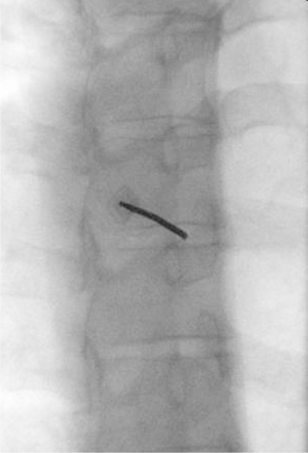
After aseptic preparation of the skin, the needle is directed in a tunneled view (Fig. 1), just lateral to the interpedicular line, keeping the needle tip medial to the costovertebral joint [9]. This is important to prevent the pleura from being penetrated. At times, the pleura may be breached even if such an approach is performed. The bent-needle technique is hence ideal. Two factors work upon the bent needle and act synergistically with the trocar to impart a medial movement on the inner needle: (1) The bend, which is oriented toward the disc; and (2) the bevel, which faces laterally, facilitate the tip to move medially upon purchasing soft tissue such as the annulus [8]. The bent-needle technique thus allows a more medial entry, just lateral to the interpedicular line and enables the needle tip to be steered laterally around the superior articular process into the herniated thoracic disc. We bend the needle approximately 1 inch from the tip with our thumbnail tangentially along the needle shaft (with the index finger under the needle) so the bevel faces away from the direction of the bend, with the trocar still engaged. The key to a good "bend" is having a smooth turn and tip in the same linear plane as the needle shaft. Care has to be taken then to ensure that the trocar can be retracted without much resistance. This is followed by the insertion of the Perc-DLG tissue ablation and coagulation SpineWand® to verify the ease of its insertion and withdrawn without weakening or fracturing the tip.
Upon contact with the intervertebral disc, the annulus will provide initial resistance and will feel "gritty" as the needle passes through it. In the thoracic intervertebral disc, this will be followed by a distinct loss of resistance as the needle enters the nucleus pulposus. The needle tip should be placed in the transitional zone between the annulus and the nucleus. This would correspond fluoroscopically to the posterior one-third of the disc space on the lateral view. The SpineWand® is then inserted and advanced until the tip of the wand is approximately 5 mm beyond the tip of the cannula, assuring that the active portion of the wand was beyond the inner layer of the annulus and in the nucleus. The depth is then confirmed in both the postero-anterior and lateral fluoroscopic views (Fig. 2). The wand is then advanced until it comes into contact with the annulus on the opposite side, which will offer some resistance. The depthstop marker on the shaft of the Perc-DLG SpineWand® is advanced close to the needle hub to designate the distal channeling limit. The process of decompression involves advancing the wand, in ablation mode, at a speed of 0.5 cm/sec and, similarly, retraction of the wand was performed in coagulation mode at a speed of 0.5 cm/sec. A total of six channels is created at the twelve, two, four, six, eight, and ten o'clock positions.
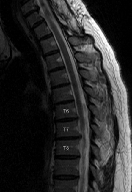
The first two patients experienced more than 75% pain relief and patient 3 experienced more than 50% pain relief. Their pain relief was sustained up to the time of the writing of this manuscript with an average of 10 months of postprocedure relief. A thoracic MRI was repeated for patient 1, one month after the procedure which showed a near complete resolution of the T6/7 and T7/8 disc prolapse (Figs. 3 and 4). However, pre and post-procedural MRI slices are not identical as they were performed in two different hospitals). Nucleoplasty of T11/12 was performed in patient 2 and the radicular left flank pain had completely resolved after the procedure along with the T11 dermatomal parasthesia. Patient 3 had incidental lower limb radicular symptoms which were not related to the T12/L1 disc prolapse. The pain over her right hip had resolved almost completely but lower limb symptoms remained. There were no reported post-operative neurological complications in all three patients.
Discussion
In their review of 280 casers, Arce and Dohrman [10] found an incidence of 75% of thoracic disc herniations below T8, with a peak incidence of 28% at T11/12. The reasons for this are due possibly to their anatomical characteristics, being thinner anteriorly and thicker posteriorly, in addition to the increased mobility in the lower segments [3]. Symptomatic herniated thoracic discs accounted for 0.25 to 0.75% of all disc ruptures and tend to present in the third to fifth decade of life, with a peak occurrence in the fourth decade [3,10].
Thoracic intervertebral discs have a well-defined nucleus pulposus, but are smaller in volumetric dimension than the lumbar disc nucleus. The nucleus pulposus are more centrally located, surrounded by a dense fibroelastic annulus fibrosus. The volumetric reduction in the thoracic nucleus pulposus, may however result in a greater degree of neural decompression due possibly to the elliptical to circular shape of thoracic discs and its relative horizontal orientation.
All three of our patients have small disc herniations with pain as their main symptom. Patients with large herniations of more than 20% or calcified herniations are unlikely to benefit from such a procedure. Disc consistency is thus an important parameter influencing treatment. For the technique of thoracic disc nucleoplasty, we recommend additional computed tomography imaging to further delineate the extent of calcification, especially when this is suspected from conventional radiographic films or T1-weighted MR images. Furthermore, we are not advocating this as a procedure to replace essential function-salvaging surgery for neurological compromise but rather as a viable option in view of its reduced morbidity in patients with mainly pain as their chief complaint, with small herniations or for patients who are otherwise unfit for conventional surgery.