Hypertension Is Related to Positive Global Sagittal Alignment: A Cross-Sectional Cohort Study
Article information
Abstract
Study Design
Cross-sectional cohort study.
Purpose
This study aimed to investigate the relationship between hypertension and spino-pelvic sagittal alignment in middle-aged and elderly individuals.
Overview of Literature
Positive global sagittal alignment is associated with poor health-related quality of life. Hypertension is associated with tissue microcirculation disorders of the skeletal muscle. We hypothesized that hypertension may be involved in positive global sagittal alignment.
Methods
In this institutional review board-approved study, 655 participants (262 men and 393 women; mean age, 72.9 years; range, 50–92 years) who underwent musculoskeletal screening in Toei town, Aichi, Japan were included. Whole spine and pelvic radiographs were taken, and radiographic parameters (thoracic kyphosis, lumbar lordosis, pelvic tilt, sacral slope, pelvic incidence, and sagittal vertical axis [SVA]) were measured using an image-analysis software. Hypertension was assessed using the standard criteria. The study participants were divided into three subgroups as per age (50–64 years, 65–74 years, and ≥75 years). We examined the differences in the radiographic parameters of those with and without hypertension in each age subgroup.
Results
In each age subgroup, there was no significant difference in the age and sex of those with and without hypertension. SVA was significantly shifted forward in the hypertension group than in the non-hypertension group in those aged 50–64 years old (32.4 mm vs. 16.0 mm, p=0.018) and in those aged 65–74 years old (42.7 mm vs. 30.6 mm, p=0.012). There was no significant difference between the hypertension and non-hypertension groups in terms of the alignment of the lumbar and thoracic spine in all the subgroups. In multivariate analysis, hypertension was a significant independent factor of forward-shifted SVA (standardized beta 0.093, p=0.015).
Conclusions
This study showed that hypertension was associated with forward-shifted global sagittal alignment.
Introduction
In recent years, with the increase in the elderly population, poor health-related quality of life (QOL) owing to spinal deformities in middle-aged and elderly people is becoming a problem [1,2]. Changes in the posture are reported to start in the 30s in women and the 50s in men [3]. In addition, previous cohort studies have reported that the proportion of people who exceed the threshold value of sagittal global alignment increases in the 60s [4]. If global sagittal alignment shifts forward, it causes deterioration of the health-related QOL [1,5]. Deterioration of the health-related QOL in middle-aged and elderly people is a very important issue because it leads to increased resource consumption for medical and long-term care. The cause of the forward shift in the sagittal global alignment is considered to be disc degeneration and osteoporotic vertebral body fracture, except for the iatrogenic flat back syndrome [6,7]. Although it was reported that disc degeneration and/or herniation are associated with spinal sagittal alignment [8,9], the root cause is yet to be identified [8]. Risk factors for disc degeneration include high blood pressure [10], impaired glucose tolerance [10,11], hyperlipidemia [10,11], and obesity [10]. These diseases are associated with arteriosclerosis [12], and not only increase the risk of cardiovascular, but also impact the musculoskeletal system due to lack of tissue microcirculation [13]. These factors may directly affect the global sagittal alignment, resulting in intervertebral disc degeneration. In fact, the impact of arteriosclerotic factors on the intervertebral disc is larger in the thoracic vertebrae that are less movable than the lumbar vertebrae [10]. Differences in the impact of arteriosclerotic factors on disc degeneration based on the site suggest that arteriosclerosis factors are indirectly related to disc degeneration. It is unclear whether these arteriosclerosis factors are actually involved in the alteration of spinal alignment; therefore, it is important to elucidate the relationship of these arteriosclerosis factors with sagittal plane alignment. We focused on hypertension, the most common arteriosclerotic factors. This study aimed to clarify whether there is a difference in the thoracic, lumbar, and global sagittal alignment in middle-aged and elderly individuals with and without hypertension.
Materials and Methods
1. Participants
This study was reviewed and approved by the Hamamatsu University School of Medicine institutional review board (IRB approval no., 15-60) and the Toei Hospital IRB (no., 201201). After obtaining approval for the research from the IRB, musculoskeletal examination and radiographic analyses were performed for people in Toei town, Aichi prefecture, Japan in 2012. Toei town has a population of 3,528 (regional official data from October 2011) and is located in the mountains. The present study, entitled the “TOEI study 2012”, was conducted as part of a musculoskeletal screening project for middle-aged and elderly individuals in Toei town. Participants aged >50 years were enrolled in this study. We obtained written informed consent from the participants for publishing their data. We investigated the following factors: age, sex, smoking status, physical exam (height, weight, body weight index, and blood pressure), radiographic examination, presence of low-back pain, health-related QOL (using the [Oswestry Disability Index [ODI] and the EuroQol 5 dimension [EQ-5D]), and history of cerebrovascular disease and heart disease [14,15].
2. Hypertension
Blood pressure was measured twice by experienced nurses, and the average value was calculated. Using the criteria of the Japanese Society of Hypertension, hypertension was defined as a systolic blood pressure of ≥140 mm Hg or a diastolic blood pressure of ≥90 mm Hg. We also defined the participants who regularly took antihypertensive medication as hypertensive.
3. Radiographic measurements
In order to evaluate spinal and pelvic alignment, standing upright full-length posteroanterior and lateral spine radiographs were taken. Radiographic films were obtained with a 1.5-m distance between the X-ray tube and the radiograph for all the participants. The standing posture was standardized; participants were asked to relax their heads while looking straight ahead without pulling in the chin and place their hands on the clavicles. The spino-pelvic parameters (thoracic kyphosis [TK, T5–T12], lumbar lordosis [LL, L1–S1], pelvic tilt [PT], sacral slope, pelvic incidence [PI], and sagittal vertical axis [SVA]) were measured twice using standard techniques [16]. The intra-and inter-observer reliability was examined using the intraclass correlation coefficient (ICC) in SVA, PT, and PI in this cohort previously. Moreover, the intra-observer ICCs for SVA, PT, and PI were 0.995, 0.996, and 0.918, respectively, and the inter-observer ICCs were 0.996, 0.990, and 0.966, respectively [17]. We excluded participants who had undergone previous spine or joint surgeries (total hip/knee replacement or femoral neck fracture surgery). Radiographic parameters that were measured with poor quality and were very difficult to assess accurately were excluded from the study. Anatomical variations with four or six lumbar vertebrae were also excluded because these factors are significantly related with the variations in the radiographic parameters.
4. Data analysis
We divided all the participants into three age subgroups (non-elderly, 50–64 years old; young-old, 65–74 years old; old-old, ≥75 years old) and examined the differences in the radiographic parameters of the hypertension group and non-hypertension group for each age group. Similarly, differences in the average age, physical parameters (height, weight, and body mass index [BMI]), and health-related QOL (ODI and EQ-5D) were also examined between the hypertension and non-hypertension groups for each age subgroup.
5. Statistical analysis
All the values are expressed as mean±standard deviation. The normal distribution of the data was evaluated using the Shapiro–Wilk test. Differences between the groups were evaluated using the unpaired two-sample t-test or Mann-Whitney test. Fisher exact test and chi-square were used to test for significant differences in the categorical study parameters between both the groups. Spearman correlations were used to evaluate the associations of SVA with age, height, and BMI. Multiple regression analysis was used to determine the independent predictors of SVA, TK, LL, and PT with age, sex, height, BMI, presence of hypertension, presence of low-back pain, and history of cerebrovascular disease and heart disease as independent variables. In the multiple regression analysis, multicollinearity was assessed as negative based on a variance inflation factor <10. A p-value <0.05 was considered statistically significant. Statistical analyses were performed using IBM SPSS statistics software ver. 24.0 (IBM Corp., Armonk, NY, USA).
Results
1. Participant background
Among 724 participants, 655 (262 men and 393 women; mean age, 72.9 years; range, 50–92 years) were enrolled in this study. Table 1 shows the age, height, weight, BMI, systolic blood pressure, diastolic blood pressure, and smoking history as per sex. The height (162.0 cm versus 148.6 cm, p<0.001), weight (60.0 kg versus 49.3 kg, p<0.001), and BMI (22.8 kg/m2 versus 22.3 kg/m2, p=0.044) of men was significantly higher than that of women. In contrast, systolic blood pressure was significantly higher in women (135.5 mm Hg versus 141.6 mm Hg, p<0.001). Smoking history was significantly more common in men (53.8% versus 2.6%, p<0.001). There was no significant sex-based difference in the prevalence of hypertension (71.0% versus 69.5%, p=0.676) or low-back pain (57.3% versus 64.5%, p=0.060) (Table 1).
2. Differences in the spino-pelvic parameters of the hypertension and non-hypertension groups
1) Non-elderly group (50–64 years old)
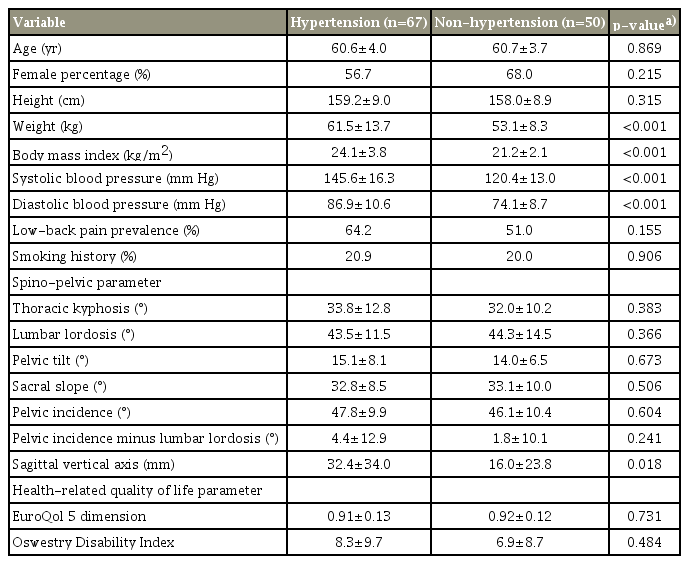
There were no statistically significant differences in the age, sex, height, prevalence of low-back pain, and smoking history of the hypertension and non-hypertension groups. Weight and BMI were significantly higher in the hypertension group. Among the spino-pelvic parameters, there was no significant difference in TK (33.8° versus 32.0°, p=0.383), LL (43.5° versus 44.3°, p=0.366), and PT (15.1° versus 14.0°, p=0.673). In contrast, the SVA was significantly shifted forward in the hypertension group (32.4 mm versus 16.0 mm, p=0.018) (Table 2).
2) Young-old group (65–74 years old)
There were no statistically significant differences in the age, sex, height, prevalence of low-back pain, and smoking history of the hypertension and non-hypertension groups. Weight and BMI were significantly higher in the hypertension group. Among the spino-pelvic parameters, there was no significant difference in TK (34.3° versus 33.3°, p=0.536), LL (41.9° versus 41.5°, p=0.849), and PT (18.1° versus 17.3°, p=0.401). However, the SVA was significantly shifted forward in the hypertension group (42.7 mm versus 30.6 mm, p=0.012) (Table 3).
3) Old-old group (≥75 years)
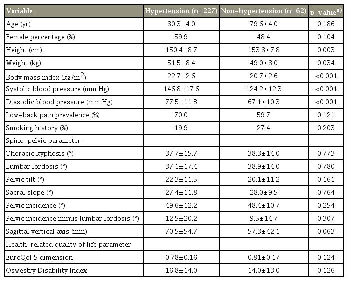
There were no statistically significant differences in the age, sex, prevalence of low-back pain, or smoking history of the hypertension and non-hypertension groups. Weight and BMI were significantly higher in the hypertension group. Those in the non-hypertension group were significantly taller. Among the spino-pelvic parameters, there was no significant difference in TK (37.7° versus 38.3°, p=0.773), LL (37.1° versus 38.9°, p=0.780), PT (22.3° versus 20.1°, p=0.161), or SVA (70.5 mm versus 57.3 mm, p=0.063) (Table 4).
3. Relationship of sagittal vertical axis and lumbar lordosis with age, sex, height, body mass index, and hypertension
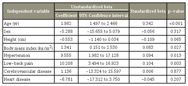
Univariate analysis showed that SVA increased with age (r=0.424, p<0.001). There was no difference in the SVA of men and women (48.3±44.6 mm versus 49.6±47.9 mm, p=0.989). Univariate analysis showed that SVA decreased with height (r=−0.169, p<0.001). SVA was not correlated with BMI (r=0.054, p=0.166) and was higher in the hypertension group than in the non-hypertension group (54.9±49.2 mm versus 35.3±36.1 mm, p<0.001). In the multivariate analysis, age (standardized beta, 0.342; p<0.001), BMI (standardized beta, 0.083; p=0.027), presence of hypertension (standardized beta, 0.094; p=0.013), and presence of low-back pain (standardized beta, 0.104; p=0.003) were significant determinants of SVA; furthermore, hypertension had an independent effect, resulting in an increased SVA of 9.5 mm (Table 5). In other multivariate analysis, only age (standardized beta, 0.128; p=0.005) was a significant determinant of TK; only age (standardized beta, −0.132; p=0.005) was a significant determinant of LL. Age (standardized beta, 0.168; p<0.001), sex (standardized beta, 0.135; p=0.011), height (standardized beta, −0.308; p<0.001), and the presence of low-back pain (standardized beta, 0.108; p=0.002) were significant determinants of PT.
4. Differences in the health-related quality of life between the hypertension and non-hypertension groups
In all the age subgroups, the EQ-5D was lower in the hypertension group, and the ODI score was higher in the hypertension group; however, these differences were not statistically significant (Tables 2–4).
Discussion
To our knowledge, this is the first study to reveal the association between hypertension and forward shift of sagittal plane alignment in middle-aged and elderly individuals. Age is closely related to sagittal plane alignment; therefore, we examined it by dividing participants into three age groups to adjust for the influence of age. Thus, we found that hypertension was significantly related to a forward shift in the global sagittal alignment in middle-aged and elderly individuals. It is noteworthy that hypertension was not significantly associated with alignment of the thoracic and lumbar spines. It is reported that the mechanism of spinal sagittal deformity involves the loss of LL due to disc degeneration or osteoporotic vertebral fracture, resulting in a sagittal plane alignment shift forward [7]. The causes of spinal sagittal deformity include disc degeneration [8] and degenerative changes in the muscles [18]; few studies have reported on the relationship of arteriosclerosisrelated diseases, such as hypertension and spinal sagittal deformity. In recent years, obesity has been reported to be associated with a deteriorating sagittal plane alignment [19,20]; however, to our knowledge, there is no report on the relationship between hypertension, a very common disease, and sagittal plane alignment. Regarding the association between degenerative musculoskeletal disease and hypertension, Yoshimura et al. [21] have reported on the association between knee osteoarthritis and hypertension in a large cohort study. Moreover, based on a cohort study, Teraguchi et al. [10] revealed that hypertension is associated with disc degeneration. However, hypertension was not related to intervertebral disc degeneration in the cervical and lumbar spines in an age-adjusted multivariate analysis; it was correlated with intervertebral disc degeneration in only the thoracic spine [10]. Hypertension is a factor strongly related to arteriosclerosis [12]. Therefore, it has been suggested that vascular insufficiency of the intervertebral disc due to arteriosclerosis might cause intervertebral disc degeneration [10]. If vascular insufficiency is involved in disc degeneration, disc degeneration will not be limited to one vertebral region. We hypothesized that hypertension produces a sagittal positive shift, resulting in intervertebral disc degeneration. The present results show that hypertension had no statistically significant effect on the local alignment of the thoracic and lumbar spine in middle-aged and elderly people aged ≥50 years; however, it did cause a forward shift of the global sagittal alignment in these subjects. This suggests that hypertension is more closely related to a positive global sagittal alignment than local disc degeneration. After adjusting for age, sex, BMI, and the prevalence of low-back pain, hypertension was an independent factor for an approximately 10-mm forward shift of the SVA. In contrast, hypertension was not a significant factor for changes in TK, LL, or PT. This supports our hypothesis that postural anomalies occur before disc degeneration in people with hypertension. In general, when the global sagittal alignment is shifted forward, the body attempts to maintain the global sagittal alignment by tilting the pelvis backwards; this is a compensatory mechanism [22,23]. However, there was no significant difference in PT between participants with and without hypertension. This indicates that pelvic compensation is more difficult in people with hypertension. The compensation mechanism occurs not only in the pelvis, but also in the lower limbs [22]; therefore, future studies can establish a more detailed mechanism by evaluating the effect of the lower limbs. Hypertension is a leading cause of both, cerebrovascular disease and heart disease [24], and they can be considered confounding factors in this research. Therefore, we performed multivariate analysis to adjust for these factors; however, only hypertension was a significant factor for positive global sagittal alignment.
Regarding the association between hypertension and positive global sagittal alignment, we created the following hypothesis. Hypertension impacts tissue microcirculation, including that in the skeletal muscle [13]. Therefore, the deteriorated microcirculation of the back muscles causes muscle fatigue and muscle degeneration that may result in posture abnormality. These effects may affect the thoracic spine, lumbar spine, and pelvis in small incremental amounts; therefore, the cumulative effect of this may be significantly related to positive global sagittal alignment. Therefore, local alignment like TK or LL may not have statistically different between hypertension group and non-hypertension group. Alternatively, the progression and development of knee osteoarthritis associated with hypertension may be involved in positive global sagittal alignment [21].
Moreover, an increased BMI was an independent risk factor for sagittal anterior shift. This result is consistent with previous reports [19,20]. In this study, we also evaluated the health-related QOL. Health-related QOL was worse in people with hypertension than in those without hypertension; however, this difference was not significant.
There are certain limitations of the present study. First, this study was a cross-sectional research. Therefore, it is not possible to clarify the cause in the relationship between hypertension and positive global sagittal alignment in this research. Second, this study involved participants of a musculoskeletal screening project conducted in a mountainous area. Therefore, the mechanical stress on the limbs and the spine may differ from those in urban areas. Third, we did not actually evaluate disc degeneration. Therefore, it is difficult to clarify whether positive global sagittal alignment affects disc degeneration or if intervertebral disc degeneration affects positive global sagittal alignment in this study. Fourth, we did not examine the sagittal alignment of the cervicothoracic junction and the lower limbs. We believe that these alignments also affect the global sagittal plane alignment; thus, these aspects require further research. To our knowledge, this is the first study to examine the relationships of atherosclerotic factors and SVA; we plan to perform a longitudinal study to clarify the manner in which hypertension is involved in positive global sagittal alignment.
Conclusions
To our knowledge, this is the first study to reveal that hypertension is related to forward-shifted global sagittal alignment in middle-aged and elderly people. In the present study, hypertension was not related with alignment of the thoracic and lumbar spines and may be more directly related to global sagittal alignment than local degeneration.
Notes
No potential conflict of interest relevant to this article was reported. DT has a donated fund laboratory by Medtronic Sofamor Danek Inc. (Memphis, TN, USA), Japan Medical Dynamic Marketing Inc. (Tokyo, Japan), and Meitoku Medical Institute Jyuzen Memorial Hospital (Hamamatsu, Japan). SO belongs to a donated fund laboratory by Medtronic Sofamor Danek Inc. (Memphis, TN, USA), Japan Medical Dynamic Marketing Inc. (Tokyo, Japan), and Meitoku Medical Institute Jyuzen Memorial Hospital (Hamamatsu, Japan).
Acknowledgements
The authors would like to express great thanks to Mr. Katsutoki Obayashi, past Mayer of Toei town, Mr. Koji Murakami, Mayer of Toei town, staff members of Municipality Health and Welfare office. The authors also would like to express great thanks to Ms. Naomi Uchiyama, Ms. Nao Kuwahara, Ms. Kumiko Yano and Mr. Taku Nagao (Department of Orthopedic Surgery, Hamamatsu University School of Medicine) for their excellent assistance of survey.


