Introduction
Detecting disc degeneration for the early diagnosis of patients with degenerative disc disease (DDD) is vital to facilitate optimal treatment, given therapies are most efficacious in the early stages of the disease. Numerous factors induce the breakdown of intervertebral discs in the spine, including osteoarthritis, disc herniation, spinal stenosis, and aging. Denervation resulting from spinal disc injury leads to muscle atrophy and loss [1]. Predicting the patientŌĆÖs disc health proximal and adjacent to the proposed surgical site is thus essential for proper surgical planning, improving functional outcomes, and abrogating the adverse effects of DDD.
To predict the severity of disc degeneration, intervertebral discs can be characterized either with T2 relaxometry to quantify the signal changes and evaluate disc health or with diffusion tensor imaging (DTI) for three-dimensional visualization [2]. The use of T2 relaxometry as a biomarker facilitates sensitive, quantitative magnetic resonance imaging (MRI) and helps identify the extent of disc damage [3,4]. Although MRI provides details of the spinal cord anatomy, conventional MRI using T2-weighted imaging is not sensitive to early morphological changes and provides no predictive quantitative biomarker profile of early degeneration. The limitations of conventional MRI also include prolonged acquisition time for multiple images.
DTI as a biomarker helps predict the severity of disc degeneration by providing qualitative information on the microstructural architecture of discs. DTI employs fiber tracking to display a ring or lamellar architecture of the annulus fibrosus (AF) that surrounds the soft inner core of the nucleus pulposus (NP). DTI also provides quantitative information using fractional anisotropy (FA) maps. The high FA values measured in the AF correlate with its highly organized, tension-resistant, fibrous collagen structure. The lower FA values measured in the NP reflect its less structured, compression-resistant, proteoglycan-rich gelatinous core [5].
The Thompson grading system is currently employed to classify the severity of intervertebral disc degeneration based on MRI into one of five stages of degeneration: stage 1 is a healthy inter vertebral disc and rest of four stages depend on the other four characteristics. Characteristics have been used to stage disc degeneration in to five stages are as follows: the number, disc height, disc fissure configuration, and distinctness of the boundary between the collagenous and the cartilaginous portions of the disc. The Thompson system was originally designed for use with anatomical slices used in experimental research [6]. Using T2-weighted images, the Pfirrmann system has shown acceptable intraobserver and interobserver reliability (0.84ŌĆō0.90 and 0.69ŌĆō0.81, respectively), with five grades of morphology and signal intensity: IŌĆōII (for apparently healthy discs of children and young adults), III (for disks with reduced T2 signal intensity), and IVŌĆōV (for disks with progressively decreased height and T2 signal) [7,8].
In this study, the extent of disc damage was determined by combining objective biomarkers using T2 relaxometry and DTI as functional tools for treating disc degeneration. We detected microstructural alterations before morphological changes and employed FA to detect morphological differences between the NP and AF, as well as degenerative changes in the NP. Finally, we propose a continuous scale with quantitative scoring to evaluate lumbar intervertebral disc degeneration.
Materials and Methods
After receiving approval from the institution review board of Vijaya Diagnostics Hyderabad (IRB approval no., 04/17/2014), we conducted an observational comparative study that included 59 patients (who provided their informed consent) and age-matched controls. All participants underwent lumbar spine MRI on a 3T MRI scanner (Philips Medical Systems, Amsterdam, The Netherlands) between 2014 and 2017. In all, we analyzed 295 healthy intervertebral discs (59 controls) and 59 patients with lumbar DDD (35 men [59.3%] and 24 women [40.6%]; male-to-female ratio, 1.4:1; age range, 29ŌĆÆ69 years; mean┬▒standard deviation age, 41.7┬▒11.80 years). We evaluated all intervertebral lumbar discs from L1 to S1 according to morphological abnormality and degeneration grades. The patient population included participants with lower back pain, sciatica, and neurological deficits. We excluded patients with spinal infections or who had undergone trauma or spinal surgery. The control and patient groups were divided into three age subgroups each: A (<30 years, n=11); B (30ŌĆÆ50 years, n=21); and C (>50 years, n=27). For each intervertebral disc, we obtained DTI-FA values for the AF and NP and T2 values for the NP in each of the three control and patient subgroups. We calculated and compared the ratios of the two parameters FA (AF/NP).
We employed the following imaging parameters: (1) 450/8 ms repetition time (TR)/echo time (TE); 372├Ś328 matrix; 33.6├Ś68.2 field of view; 4-mm slice thickness; 1.0-mm slice gap and one number of excitations (NEX) for T1 sagittal view; (2) 2,709/100 ms TR/TE; 336├Ś255 matrix; 32.3├Ś57.7 field of view; 4-mm slice thickness; 0.4-mm slice gap and 2 NEX for T2 at the intervertebral disc levels; (3) 6,248/60 ms TR/TE; 2-mm slice thickness; 0.5-mm slice gap; 2 number of signal averages; 112├Ś112 matrix; 29.8 bandwidth; b-values of 0 and 800; 15 directions for DTI in sagittal; and (4) 15, 30, 60, 80, 90, 120 ms TE for T2 maps.
1. Data analysis
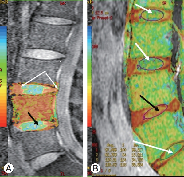
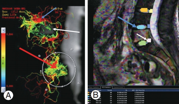
We generated FA maps using Fibre Track software (Philips Healthcare, BEST, The Netherlands) provided with the MRI equipment. FA maps of the AF and NP were coŌĆæregistered with T2 images. We analyzed the T2 relaxometry maps by region of interest (ROI) in the NP at five disc levels using T2 Map Software (IntelliSpace Portal workstation, Philips Healthcare), which generated color-coded T2 maps. ROIs were placed in the NP, avoiding end-plate regions and the disc periphery to generate FA (AF/NP) ratios and T2 values for the NP. We calculated the T2 values and FA (AF/NP) ratios for the control and patient groups for the statistical analysis.
2. Statistical analysis
The T2 values and FA (AF)/FA (NP) ratios for the control and patient groups were calculated for the statistical analysis using PearsonŌĆÖs correlation coefficient. We employed SPSS for Windows ver. 17.0 (SPSS Inc., Chicago, IL, USA) for the statistical analysis and performed the following correlations for both groups: age versus FA (AF/NP) ratio and T2 map versus FA (AF/ NP) ratio.
We evaluated the degree of correlation using the r absolute value as follows: r>0.6 was defined as strong; 0.5< r Ōēż0.6 as moderate; and rŌēż0.3 as weak. Statistical significance was set at p<0.001.
Results
We categorized the MRIs of 590 intervertebral discs of 118 participants (59 healthy volunteers and 59 patients). Of these, 566 discs were successfully measured on T2 relaxometry images (Fig. 1A, B) and FA maps (Fig. 2A, B).
We excluded 24 discs due to highly reduced disc height resulting from degeneration, those where an optimal ROI (n=17) was placed at L4ŌĆōL5 and L5ŌĆōS1 and those obscured due to susceptibility artifacts (n=7).
Among the healthy controls, subgroup A had a higher T2 and AF/NP ratio than subgroup B; however, the AF was not significantly different. Subgroups A and B had a higher T2 and AF/NP ratio than subgroup C but lower NP and AF values (Table 1).
Among the patients, there were no differences between subgroups B and C. Subgroup A showed higher T2 values than subgroup B and had a higher T2 and FA (NP) ratio than subgroup C (Table 2).
1. Healthy controls versus patients
Subgroup A of the healthy controls had a higher T2 and AF/NP ratio but lower NP than subgroup A of the patients. Subgroup B of the healthy controls had a higher T2 and AF/NP ratio but lower NP and AF values than subgroup B of the patients. Subgroup C of the healthy controls had a higher T2 and AF/NP ratio but lower FA (NP) values than subgroup C of the patients.
2. Correlations
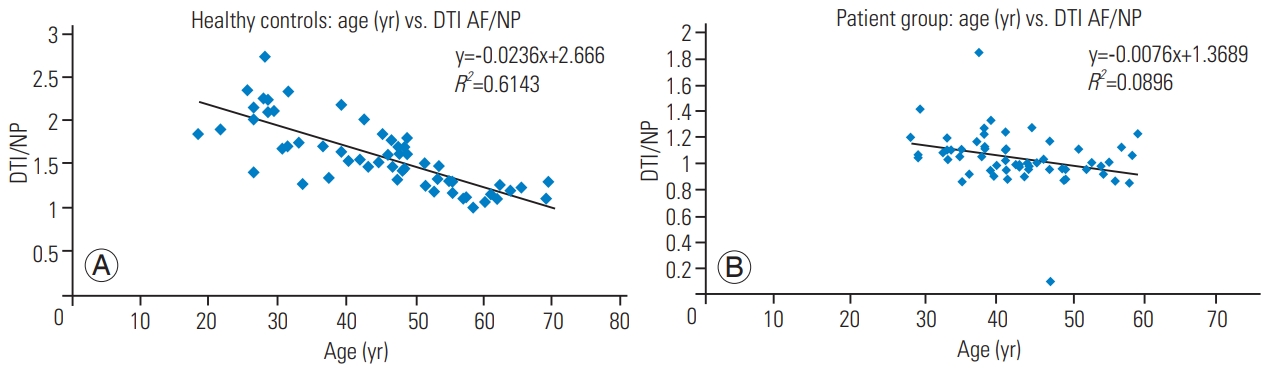
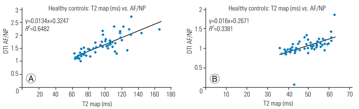
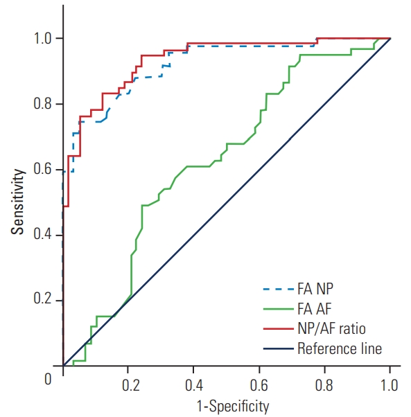
AF/NP values were strongly correlated with age and with the T2 map (ms) in the healthy controls but not in the patients. In the healthy volunteers, there was no correlation between R2 values <0.300 and FA (AF/NP), and there was a strong increasing correlation with increasing R2 values >0.300 and a trend for R2 values >0.600. AF/NP was strongly correlated with increasing age in the healthy controls but not in the patients (Fig. 3A, B). When comparing the T2 map versus AF/NP for the healthy volunteers and patient group,there was no correlation with R2 values <0.300, while there was a strong increasing correlation with increasing R2 values >0.300 and a trend for R2 values >0.600 (Fig. 4A, B). The receiver operator characteristic curves showed an area under the curve of Ōēź0.700, which was considered significant. Both FA (NP) and NP/AF ratios are reportable and significant. For an optimum cutoff value of 0.509, we observed a sensitivity of 98% and specificity of 81.5% (Fig. 5).
The control group presented mean T2 values >120 ms in subgroup A, 90ŌĆÆ100 ms in subgroup B and approximately 70 ms in subgroup C (p<0.001). The patient group presented mean T2 values of 55.8┬▒4.4 ms in subgroup A, 48.5┬▒6.9 ms in subgroup B and 45.8┬▒8.7 ms in subgroup C (p<0.050). These results indicate a significant difference in T2 values in degenerated discs in patient subgroups A and B compared with the normal healthy controls in the corresponding subgroups.
Similarly, the DTI-FA values for the NP and AF expressed as a ratio (FA AF/NP) showed significant statistical differences (p<0.001), indicated by higher FA values in the NP and lower FA (AF/NP) ratios in the patient population than in the healthy controls in subgroups A and B. T2 values and DTI indices for subgroup C (controls and patients) were not statistically significant due to age-related microstructural changes in the NP, especially in subgroup C of the healthy controls, given that differences in the FA values for NP and AF are not specific to degeneration alone.
The T2 relaxometry scoring tool provides potential benefits for quantitatively grading the early stages of DDD, wherein T2 values decrease linearly with increased degeneration. DTI-FA values can characterize the amorphous NP showing low FA values and differentiate it from AF having high FA in a healthy disc. In a degenerated disc, the NP shows increased FA values. As the degeneration advances, the NP presents characteristics similar to the AF, including fibers in the fiber tracking. The FA (AF/NP) ratio can therefore act as a potential biomarker.
3. Predictive disc health score
We therefore propose the following simple predictive disc health scoring system based on T2 relaxometry and DTI indices.
H (healthy): T2 values >120 ms, FA (NP/AF) ratio >2, mean FA (NP) between 0.4 and 0.55.
DE (early degeneration): T2 values <100 ms, FA (NP/AF) ratio <1.5, mean FA (NP) between 0.6 and 0.65.
DA (advanced degeneration): T2 values <80 ms, FA (AF/NP) ratio <1.2, mean FA (NP) between 0.8 and 0.85.
Discussion
The present study examined the potential of combining biomarkers using T2 relaxometry and DTI to characterize intervertebral disc health in various age groups. The intervertebral disc is a fibrocartilaginous joint that supports spinal forces, facilitating joint movement. The disc consists of tension-resistant AF and compression-resistant NP, which together mechanically stabilize the disc [2]. Healthy intervertebral discs show high T2 signal intensity, while degenerated discs present lower T2 signal intensity. The criteria for classifying intervertebral disc degeneration are therefore based on inspecting T2-weighted images. MRI findings of disc degeneration include low T2 signal intensity, reduced height and annular fissures in the disc.
1. T2 relaxometry
The decay constant for MRI T2 signal intensity, known as the T2 relaxation time, is an intrinsic tissue property that reflects the discŌĆÖs molecular framework (consisting of water, proteins, collagen, and other solutes) and can be calculated from a series of images obtained with different echoes and recovery times or by relaxometry [4,7]. T2-weighted multiecho sequences provide T2 measurements in approximately 6 minutes with a 1.5-T system and in approximately 8 minutes on a 3T system with a 6-echo sequence [9]. Mwale et al. [10] reported that quantitative MRI indices (relaxation, magnetization transfer ratio, and apparent diffusion coefficient [ADC]) are driven by the disc matrix composition (water, proteoglycan, and collagen) and matrix integrity as represented by collagen denaturation. Different regions within the disc demonstrate varying signal intensity and T2 relaxation values. While the intranuclear cleft and the peripheral annulus show reduced T2 values due to their high fiber content, the NP shows higher T2 values due to its amorphous nature [11]. Diurnal variations in water content due to degeneration can be measured with T2 relaxometry [11-16].
2. Diffusion tensor imaging and disc disease
DTI illustrates the intervertebral disc microstructure based on diffusion-driven molecular mobility through diffusion tensor calculation. Diffusion represented by the ADC as a biomarker decreases in degenerated discs, correlating in a direction-dependent manner with intradiscal water and disc matrix integrity. ADC values might help with evaluating disc degeneration due to their sensitivity to water content and the geometrical packing of the collagen network structure [17]. Given that it is not truly quantitative, ADC characterizes a single scalar coefficient and is dependent on the number of directions used for measurements. Invariant indices are necessary for an accurate depiction of the intervertebral disc microstructure [18-21].
We observed a mean decrease in FA values in subgroups A, B, and C, with progressive degeneration. The NP has a high molecular weight with fixed negative charges and thereby induces osmosis [22]. The AF is composed of type I collagen fibers in the lamellae and limits the swelling of the NP and adjacent vertebral segmental motion. The swelling pressure of the NP is opposed by tensile forces in the collagen fibers of the AF [3,23]. Physiological degeneration of lumbar intervertebral discs usually starts in the second decade of life. The metabolic derangement of NP involves decreased water content, progressive loss of proteoglycan and damage to the collagen matrix [24,25]. Our data indicates that the use of T2 relaxometry provides a qualitative and quantitative diagnosis of DDD. Conventional MRI has acceptable qualitative accuracy in detecting morphological changes as part of the diagnostic characterization of disc disease due to aging and degeneration but is unable to identify the clinical significance in asymptomatic individuals [26,27]. Recent studies have suggested that MRI cannot provide information on the cause of back pain in DDD, given that similar morphological changes have been observed in patients with back pain and in asymptomatic individuals [6,28-30].
The relaxation times of the NP change by approximately 10% per decade in normal disks. A 20%ŌĆō50% change has been shown in Pfirrmann grade III degeneration. T2 relaxation times in the intervertebral disc have been shown to decrease gradually with age and degeneration as a result of diminishing water and glycosaminoglycan content in the disc [1,15]. Given that T2 correlates very closely with water content, T2 in theory could provide a continuous measure of hydration in the discŌĆÖs cartilage matrix.
The increased FA in the NP, in contrast to the unchanged FA in the AF of degenerated discs, indicates multiple changes in the NP and the AF that may not correlate with morphological changes seen in T2W images. Chan et al. [18] demonstrated that mean FA values in the AF were significantly higher than those in the NP and observed that mean FA values in the NP increased as the degeneration progressed from the early to advanced stage, with a relatively inert change in mean FA values in the AF. The authors concluded that early disc degeneration could be detected by including the quantitative DTI indices in the clinical imaging protocol [5,18].
The disadvantages of current grading scales are their subjectivity, a lack of specific criteria for characterizing distinct or indistinct boundaries and their ambiguity regarding degeneration versus age-related changes. These scales are discontinuous, rendering only obvious measurable changes in disc morphology and providing limited value in the short-term monitoring of the intervertebral disc signal. These scales are also unable to distinguish the progression of degeneration and repair without a major morphological change. Disc degeneration or regeneration is also not effectively demonstrated with discontinuous scales such as the Thompson and Pfirrmann scales.
A continuous scale or scoring system is therefore required to evaluate intervertebral disc disease [4,7]. Our scoring system is both qualitative and quantitative, using T2 relaxometry color maps and values for disc hydration and water content. FA values from the NP and AF and their ratio provide a structural correlation. This study is not only predictive but is also potentially reproducible.
Conclusions
This study shows that quantitative MRI with T2 maps and DTI provides objective evidence of disc degeneration (early/advanced) and facilitates the use of novel diagnostic quantitative imaging therapies for the early detection of disc degeneration. This method also helps predict the disc health of adjacent segments above and below the level of the proposed surgery or instrumentation. Given that instrumentation accelerates disc degeneration by increasing the loading on noninstrumented discs, it is vital to predict which disc adjacent to the proposed fusion level will degenerate early. Aging and disc tissue degeneration correlate with the transformation of the NP fluid-like structure to the cartilaginous fibrous structure (AF) scored by T2 relaxometry and DTI. This scale is relatively simple, practical, and objective and helps provide guidance on the level of surgery in avoiding unnecessary long segment fusions with short- and/or long-term complications.