Asian Spine J Search

CLOSE


Asian Spine J > Volume 15(3); 2021 > Article
Luzzi, Gragnaniello, Marasco, Lucifero, Del Maestro, Bellantoni, and Galzio: Subaxial Vertebral Artery Rotational Occlusion Syndrome: An Overview of Clinical Aspects, Diagnostic Work-Up, and Surgical Management

Abstract

Extrinsic compression of the subaxial vertebral artery (VA) may cause rotational occlusion syndrome (ROS) and contribute to vertebrobasilar insufficiency potentially leading to symptoms and in severe cases, to posterior circulation strokes. The present literature review aimed to report the main clinical findings, diagnostic work-up, and surgical management of the subaxial VA-ROS, the diagnosis of which can be difficult and is often underestimated. An illustrative case is also presented. A thorough literature search was conducted to retrieve manuscripts that have discussed the etiology, diagnosis, and treatment of ROS. Total 41 articles were selected based on the best match and relevance and mainly involved case reports and small cases series. The male/female ratio and average age were 2.6 and 55.6±11 years, respectively. Dizziness, visual disturbances, and syncope were the most frequent symptoms in order of frequency, while C5 and C6 were the most affected levels. Osteophytes were the cause in >46.2% of cases. Dynamic VA catheter-based angiography was the gold standard for diagnosis along with computed tomography angiography. Except in older patients and those with prohibitive comorbidities, anterior decompressive surgery was always performed, mostly with complete recovery, and zero morbidity and mortality. A careful neurological evaluation and dynamic angiographic studies are crucial for the diagnosis of subaxial VA-ROS. Anterior decompression of the VA is the cure of this syndrome in almost all cases.

Introduction

Subaxial vertebral artery (VA) rotational occlusion syndrome (ROS) causes a transient or permanent decrease in blood flow in the posterior cerebral circulation that is associated with the occurrence of dizziness, drop attacks, visual disturbances, sensitive and motor symptoms, and even devastating strokes. These symptoms are classically triggered by the axial rotation of the head within the physiologic range of motion [1].
Disk herniation, spondylosis, ligamentous hypertrophy, bony defects, and abnormalities in the course of the VA across the transverse foramina are the main etiological factors. Atherosclerosis worsens the frequency and severity of the symptoms [2-9]. The treatment of the subaxial VA-ROS is elective surgical that aims to decompress the VA and restore normal blood flow in the posterior circulation [10-15]. Nevertheless, literature about the overall management of this syndrome is limited and mostly consists of case reports or small case series [2,3,6,16-54].
This literature review addresses the clinical aspects, diagnosis, and surgical management of subaxial VA-ROS. An illustrative case is also reported.

Methods

Two separate systematic reviews of the literature were conducted using the Ovid Medline and EMBASE databases to identify articles relevant to the natural history, diagnosis methods, treatment modalities, and outcomes of patients affected by VA-ROS. A population, intervention, comparison, and outcome search strategy was used for each review. Keyword and MeSH search terms were used including “axis, cervical vertebra,” “vertebral artery,” “occlusion syndrome,” “cervical vertebra,” “compression syndrome,” “bow hunter’s syndrome,” “decompression,” “dynamic angiography,” “vertebrobasilar insufficiency,” and “head movements.” All the manuscripts published in English were eligible for inclusion. This search strategy selected articles that described the diagnosis, natural history, and treatment of the VA-ROS. Reference lists of the included manuscripts were studied to identify additional relevant publications. Further articles were included following a manual search of the literature.

Results

1. Literature volume

The search retrieved 77 articles. After applying the exclusion criteria and removing duplicates, 41 relevant articles were selected and have been reported in Table 1.

2. Demographic and clinical data

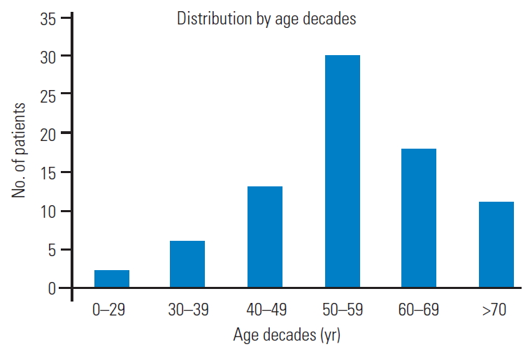
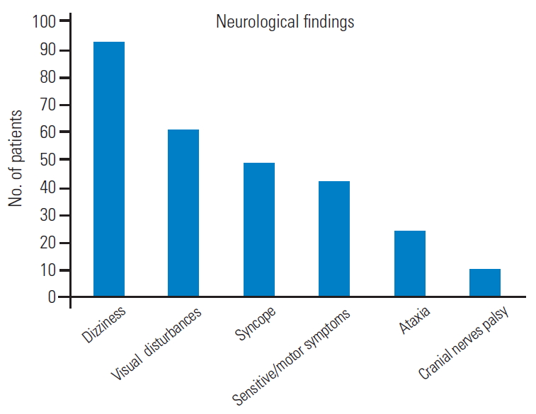
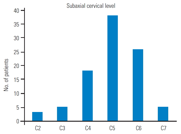
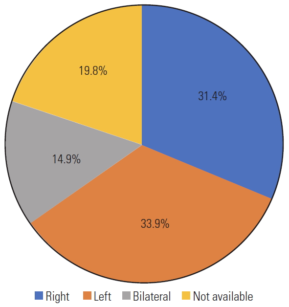
Total 121 patients were included in the study. The male/female ratio was 2.6 although sex was not reported in 24 patients. The average patient age was 55.6±11 years, and those in the fifth decade were more affected. Graph 1 shows the distribution by age decades (Fig. 1). In cases with vascular risk factors for stroke, the average frequency of hypertension, diabetes mellitus, and hyperlipidemia were 66%, 19%, and 15%, respectively (Fig. 2). Dizziness, visual disturbances, and syncope had a prevalence rate of 76.8%, 50.4%, and 40.4%, respectively (Fig. 3). The C5 level was the most affected (31.4%) followed by the C6 level (21.4%) (Fig. 4). Left side had prevalence (33.9% versus 31.4%) and in 14.9% of the cases, the cause of occlusion involved both the sides (Fig. 5).

3. Etiological factors

Cervical spondylosis was the most common causative factor (46.2%), followed by muscle band (33.8%), fibrous perivascular ring (21.4%), and disk herniations (7.4%). Less common reasons were degenerative spondylolisthesis (2.4%) and biomechanical traumatic instability (1.6%) (Fig. 6).

4. Diagnostic work-up

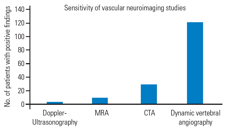
Diagnostic work-up involves different neurovascular imaging techniques whose sensitivity widely varies by type. Dynamic VA angiography, initially non-subtracted and then subtracted (digital subtraction angiography, DSA), has been crucial in the diagnosis in 100% of the reported cases. Computed tomography (CT) and magnetic resonance angiography were performed in 24.7% and 8.2% of the cases, respectively. Doppler ultrasound (DUS) standalone allowed the diagnosis in only four cases (3.3%) [24-26,32] (Fig. 7).

5. Indication for surgery

Considering the neuroimaging evidence of VA-ROS, the indication for surgery was based in all cases on the severity of the symptoms, negative impact on the performance of normal daily and working activities, and failure of conservative therapies. Conversely, conservative treatment in collar brace that also involved the establishment of an antiplatelet or anticoagulant therapy, to be decided case-bycase based on the coexistence of vascular risk factors for stroke, may have a rationale in elderly patients with minor symptoms.

6. Choice of the approach and surgical corridors

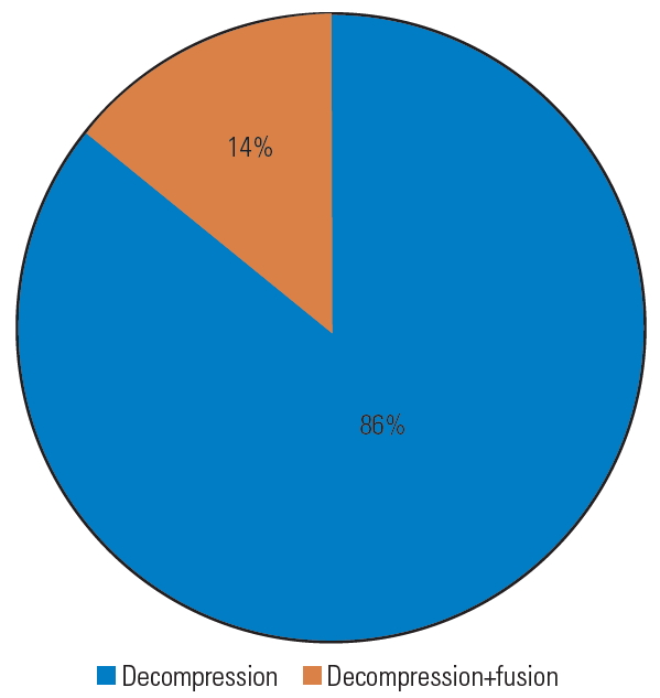
In all cases, anterior decompression of the VA was performed, accompanied by fusion in 16% of the cases (Fig. 8). An anterior Smith–Robinson (Baley–Cloward) approach was used in all cases with the exception of the series by George and Bruneau, where an anterolateral presternocleidomastoid retro-jugular route was reported [11,55-59].

7. Complications

No complications attributable to the surgical treatment have been reported, except in a case where the occurrence of Horner’s syndrome suggested injury to the sympathetic chain [23].

8. Overall outcome

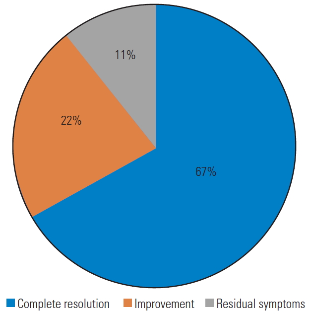
In 67% of the cases, the pre-operative symptoms disappeared immediately after surgery, and vascular neuroimaging revealed a full release of the VA with no residual signs of extrinsic compression. Further, 22% of the patients showed significant clinical improvement, while 11% reported only partial neurological amelioration with residual symptoms (Fig. 9). No specific scales were used for this evaluation.

9. Illustrative case

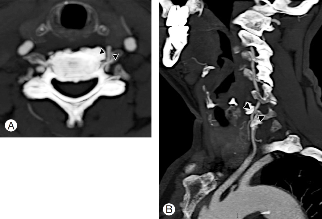
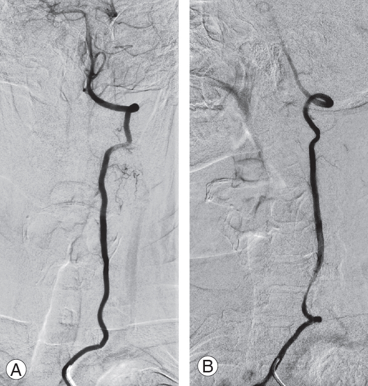
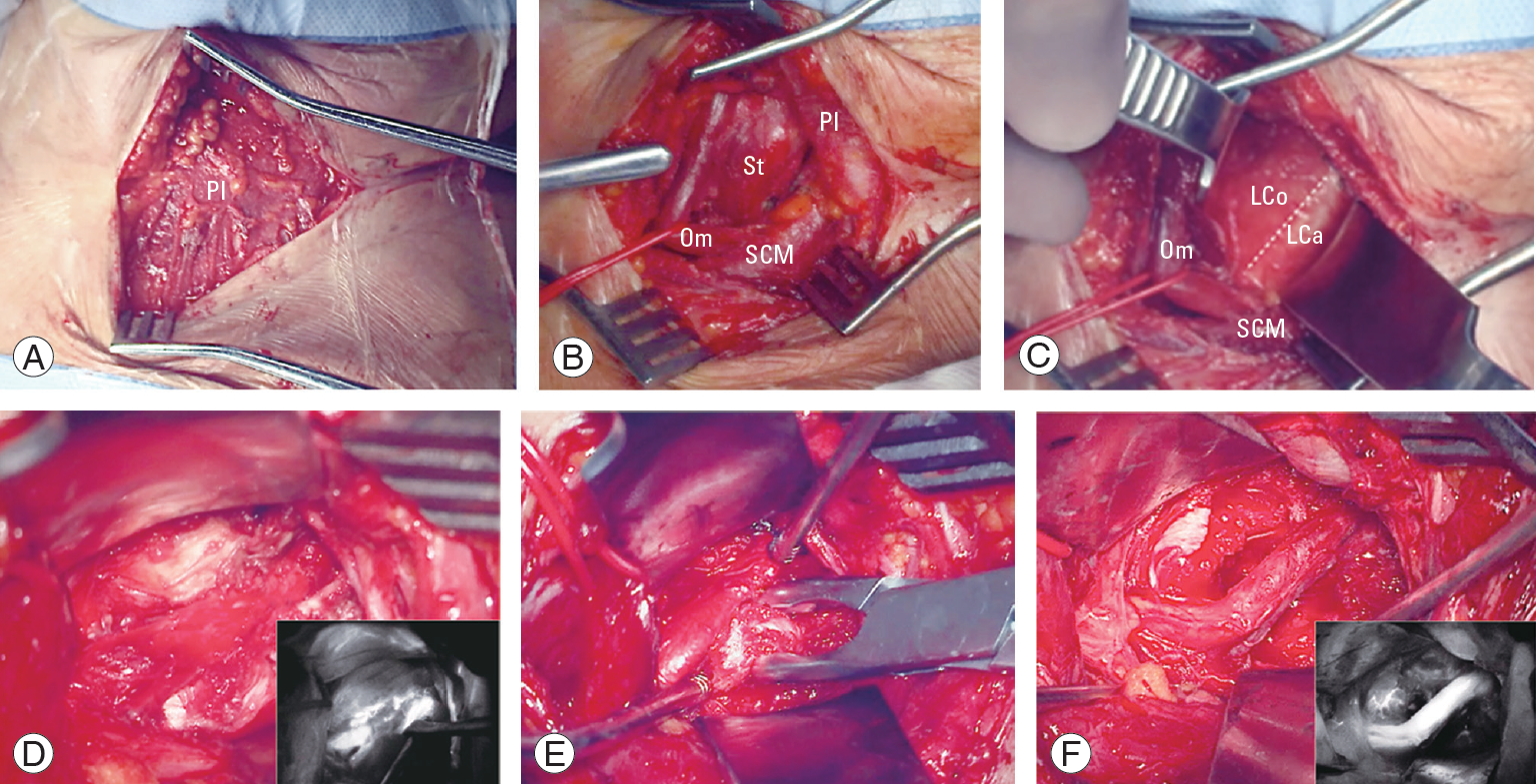
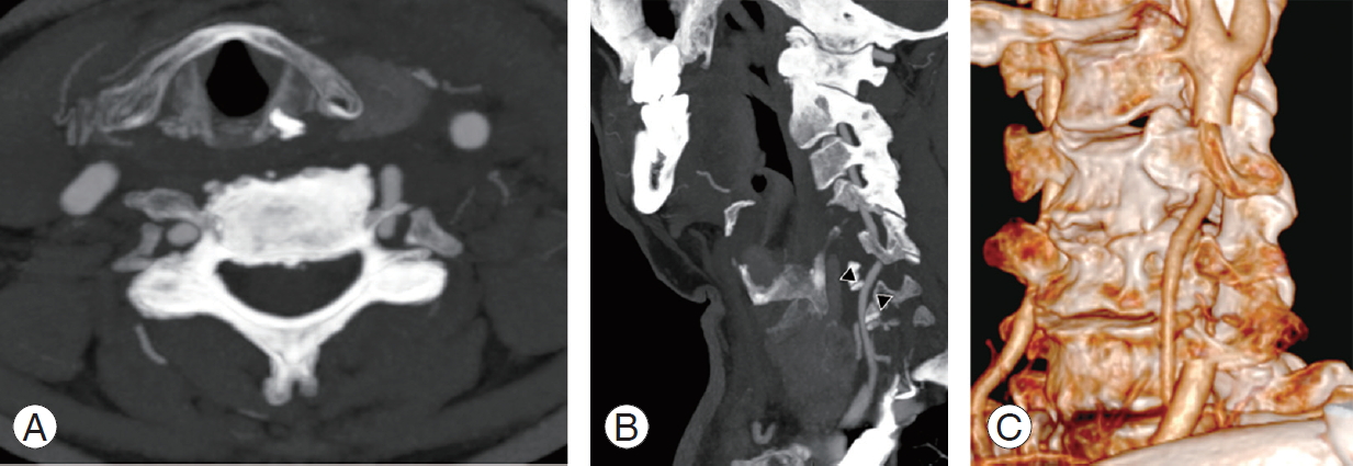
A 74-year-old male patient with medical history of hypertension experienced seven consecutive drop attacks in duration of 14 months. Cardiovascular investigations, blood tests for thrombophilia, and DUS were all within normal limits. The last attack was in association with left facial nerve palsy, rotatory nystagmus, dizziness, and numbness in the right arm. The CT and magnetic resonance imaging findings were normal; therefore, a transitory ischemic attack was suspected, and the patients underwent systemic thrombolysis, followed by anticoagulation with a factor Xa inhibitor, rivaroxaban. As the drop attacks manifested again in the 3 months after initiating anticoagulation therapy, the patients came to our attention. A comprehensive neurological examination revealed an association between the right axial rotation of the head >45° and early appearance of nausea, blurred vision, and dizziness. CT angiography revealed dynamic tethering of the VA at C5–C6 during right axial rotation of the head caused by a C6 osteophyte of the left uncinate process narrowing the anterior ramus of the satellite transverse foramen (Fig. 10). Catheter-based dynamic angiography confirmed these findings (Fig. 11). The patient underwent selective unroofing of the left C6 transverse foramen trough an anterolateral approach. The retro-longus colli corridor allowed easy reach and exposure of the subaxial portion of the V2 segment of the VA (Fig. 12). Postoperative static and dynamic CT and catheter-based angiography confirmed complete release of the VA (Fig. 13). The drop attacks subsided. Anticoagulant therapy was maintained for 1 month postoperatively and then discontinued.

Discussion

The present comprehensive literature review about the management of the subaxial VA-ROS showed that most of the publications were case reports or small cases series. Dynamic angiography is the gold standard for this challenging diagnosis, and decompressive surgery performed via the anterior or anterolateral access route can be curative.
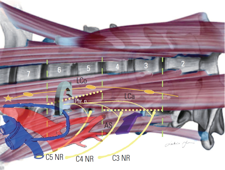
Based on a literature review, the biggest challenge concerning VA-ROS is the delay in diagnosis owing to the dynamic and intermittent nature of artery compression, thus often mimicking cardiovascular or other neurovascular pathologies. The transient symptomatology is caused by the axial rotation of the head during a careful neurologic evaluation. George et al. [60] reported that, paradoxically, chronic, static, and concentric causes of compressions, as those secondary to tumors abutting and slowly compressing the VA, are better tolerated because collateral muscular vascular supply develops to vicariate even occlusions of the dominant VA. From the clinical standpoint, compressions of the V2 segment are generally better tolerated than those of V1 and V3 owing to the protective role played by the transverse processes. V1 is susceptible to compressions by fibrous bands and muscle fibers [13,22,60], while V3 is the most commonly affected segment as pathology at the CV junction and C1–C2 is far more common [60].
In an extensive literature review on the pathophysiology and diagnosis of vertebrobasilar insufficiency, Lima Neto et al. [61] reported that this syndrome is more commonly observed in men after the fourth decade of life. Similar data have been reported by Savitz and Caplan [1].
Vertebrobasilar insufficiency is linked with three different mechanisms, including blood flow drop, embolic events, and autoregulation loss [3,60,62]. In particular, for the VA-ROS involving V2 in the subaxial cervical spine, osteophytes are the most common cause, and the embolization from thrombotic material secondary to a chronic sub adventitial mechanical stress seems responsible for the ischemic events [60].
Atherosclerosis is the most important coexisting condition favoring this syndrome; smoking, hypertension, hyperlipidemia, diabetes, and obesity are considered risk factors [61]. Not surprisingly, atrial fibrillation, infective endocarditis, and systemic hypercoagulable state further increase the stroke risk in VA-ROS patients.
Hence, the rationale of antiplatelet/anticoagulant regimen in those patients that not surgical candidates. Machaly et al. [63] demonstrated that vertigo is significantly more common in patients with severe forms of cervical spondylosis, while Olszewski et al. [64] documented a reduction in blood flow in the basilar artery and VAs that could be attributed to the extrinsic compression caused by osteophytes. Bulsara et al. [3] clarified that uncovertebral joint osteophytes cause symptoms during axial rotation and extension of the head, respectively.
Regarding those forms sustained by muscle and fibrous perivascular bands, male and younger patients are at a higher risk, with any probability because of the frequent muscular hypertonia.
Our data highlighted a major involvement of the C5 and C6 levels, similarly to that reported by Nagashima [19]. This subgroup of patients involved mostly men, and risk factors for atherosclerosis were present in all cases, thus confirming the aforementioned theory.
Labyrinthic symptoms associated with vertebrobasilar insufficiency are reported to be mainly caused by the lack of collateral vascular supply of the labyrinthine branches that are tiny and terminal in case of impairment of the vertebrobasilar arterial system [1,61,65]. Moubayed and Saliba [66] also postulated a possible direct ischemia of the labyrinth, secondary to peripheral hypoperfusion or ischemia.
The sensitivity of different neurovascular imaging techniques in the diagnosis of the subaxial VA-ROS warrants discussion. Although simple, non-invasive, repeatable and cost-effective, DUS had the worse sensitivity in our study. However, although it had a sensitivity lower than that of catheter-based angiography, dynamic CT angiography allows direct visualization of the bony compression on the VA, as well as its postoperative release. With a sensitivity and a specificity of 100%, dynamic DSA remains the gold standard for the radiological diagnosis of the subaxial VAROS, allowing detailed appreciation of compression, but also giving information about vascular dominancy of one of the VAs, size of the posterior communicating arteries, anatomical variations of the VA at its termination in the posterior-inferior cerebellar artery, and existence of collaterals. With respect to the sensitivity and specificity of dynamic DSA, our data are consistent with those reported in the literature. It is noteworthy that Toole and Tucker [67] reported that in case of VAs that are different in size, the ischemic event may be attributable to the compression of the non-dominant VA after head rotation to the contralateral side. Selective catheterization of the VAs is obviously of utmost importance to confirm the diagnosis and for surgical planning.
Stand-alone surgical decompression, achieved with selective unroofing of the V2 segment of the VA at the transverse foramen, can be curative for subaxial VA-ROS of the V2 segment, if dynamic compression is demonstrated. In fact, our review demonstrated that the cumulative rate of complete recovery/improvement of symptoms is 89%. For the remaining 11% of the patients with residual symptoms, we conducted an additional different decompressive technique for the removal of the so-called “fibrous band” if further impingement was noted at the V1 segment proximal to the artery entering the foramen at C6 [60].
The addition of a posterior instrumented fusion is seldom necessary and is necessary only if instability is noted or the compression is due to other causes, such as a disc herniation or spondylolisthesis [11,24,25,34,44,48].
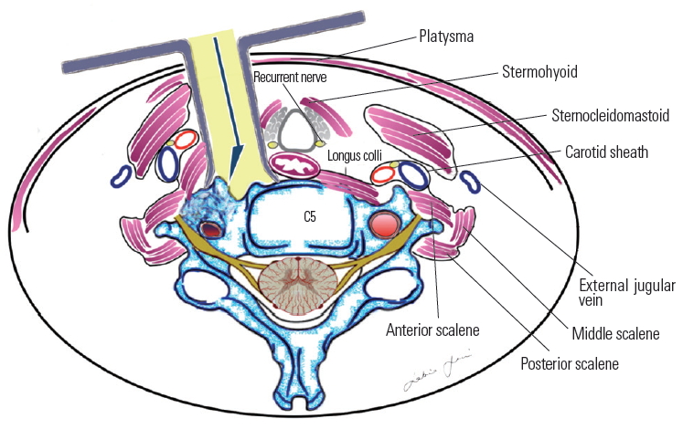
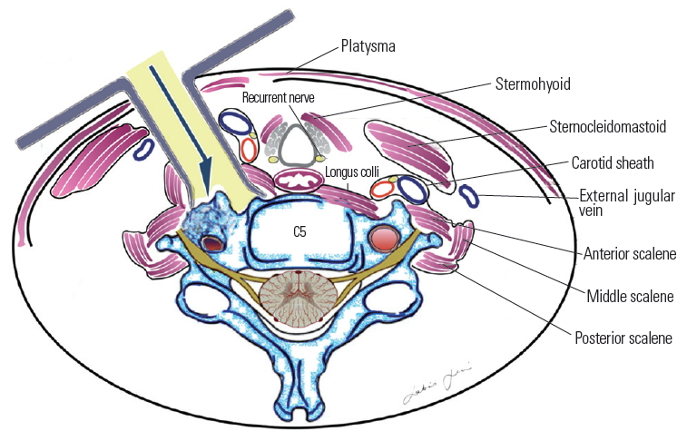
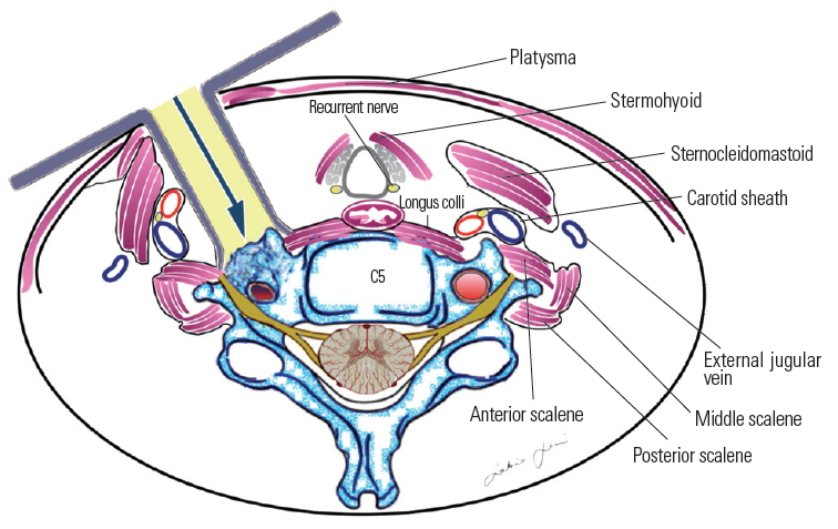
Most authors recommend simple lateral extension of the classic Smith–Robinson (Bailey–Cloward) technique [68-70] as suitable to expose the prevertebral musculature and VA [13,14]. This represents a routinely performed operation with which most neurosurgeons are familiar. The approach entails subperiosteal dissection of the longus colli muscle and the exposure of the anterior tubercle of the transverse foramen (Fig. 14). This approach is considered an anterior access route. An anterolateral approach was instead popularized by Bruneau et al. [10-12,57] to obtain an easier and more direct access to the subaxial V2 segment of the VA. The anterolateral approach involves a pre-sternocleidomastoid retro-jugular exposure of the prevertebral area along with the transection of the longus colli muscle (Fig. 15). As we report here in this illustrative case, we prefer the anterolateral approach for decompression of the subaxial VA with some modifications, including the precarotid exposure of the prevertebral muscles. The rationale of the precarotid exposure lies in the fact that injuries of the thoracic duct are very uncommon through this route because it has a more lateral course at the C4–C6 level [10,71-77] (Fig. 16).
Our personal experience also led us to delineated a retro-longus colli and a pre-scalenic prevertebral corridor for C5–C6 and C3–C4 level, respectively, to avoid injuries of the sympathetic chain and phrenic nerve, here having an upward and lateral-ward course [78-80] (Fig. 17).
Having the same advantages already reported by our group for aneurysms and arteriovenous malformations, intraoperative indocyanine green video angiography and microdoppler ultrasonography are also useful adjuncts [81-88].

Conclusions

Subaxial VA-ROS classically causes drop attacks, dizziness, and visual disturbances triggered by axial rotation or head extension. A careful neurological evaluation is paramount to suspect the syndrome. Compressions of the V2 segment of the VA are caused in most cases by osteophytes that involve the uncoapophyseal joints at the C5 and C6 level. Compressions of the V1 segment of the artery relate to the presence of fibrous bands and muscular compression. Dynamic compressions of the VA by tumors are less common.
Dynamic DSA is the gold standard for establishing a diagnosis although dynamic CT angiography is the first step to delineate the extrinsic causes of compression. Anterior decompression of the subaxial VA affected segment is curative in most cases.

Conflict of Interest

No potential conflict of interest relevant to this article was reported.

Notes

Author Contributions

Conceptualization and writing–original draft preparation: Sabino Luzzi, Cristian Gragnaniello; soft ware and valida-software and validation: Stefano Marasco, Alice Giotta Lucifero; review and editing: Mattia Del Maestro; validation: Giuseppe Bellantoni; and supervision: Renato Galzio.

Acknowledgments

We want to thank Eng. Giorgia Di Giusto for her outstanding and valuable support in formatting the manuscript and editing the graphs and figures.

Fig. 1.
Bar graph showing the distribution by age decades.
asj-2020-0275f1.jpg
Fig. 2.
Pie graph reporting the prevalence of the main vascular risk factors for stroke.
asj-2020-0275f2.jpg
Fig. 3.
Bar graph showing the main neurological findings.
asj-2020-0275f3.jpg
Fig. 4.
Bar graph describing the rate of the involvement of the different subaxial cervical levels.
asj-2020-0275f4.jpg
Fig. 5.
Pie graph about the sideness.
asj-2020-0275f5.jpg
Fig. 6.
Bar graph showing the prevalence of the different etiological factors.
asj-2020-0275f6.jpg
Fig. 7.
Bar graph reporting the sensitivity of the vascular neuroimaging techniques. MRA, magnetic resonance angiography; CTA, computed tomography angiography.
asj-2020-0275f7.jpg
Fig. 8.
Pie graph about the type of surgical treatment.
asj-2020-0275f8.jpg
Fig. 9.
Pie graph reporting the overall outcome.
asj-2020-0275f9.jpg
Fig. 10.
Axial (A) and sagittal (B) contrast enhanced CT angiography showing a left C5–C6 margin-somatic osteophyte narrowing the near transverse foramen and causing a severe compression of the vertebral artery (arrowheads).
asj-2020-0275f10.jpg
Fig. 11.
Catheter-based digital subtraction angiography of the left vertebral artery. Static anterior-posterior projection (A) and dynamic right oblique projection (B) acquired during the right axial rotation of the head, and revealing a tethering of the vertebral artery at C6 level.
asj-2020-0275f11.jpg
Fig. 12.
Main surgical steps of the left anterolateral approach to the sub-axial V2 segment of the vertebral artery. (A) Platysma muscle; (B) pre-sternocleidomastoid precarotid corridor with medialization of the omohyoid muscle; (C) identification of the retro-longus colli corridor; (D) picture-in-picture operative image showing the compression of the vertebral artery by osteophyte and the indocyanine green video angiography (IR 800, Zeiss Kinevo 900; Carl Zeiss AG, Oberkochen, Germany); (E) unroofing of the left C6 transverse foramen. (F) Picture-in-picture operative picture and video angiography showing the vertebral artery completely decompressed. Pl, platysma muscle; St, sternothyroid muscle; Om, omohyoid muscle; SCM, sternocleidomastoid muscle; LCo, longus colli muscle; LCa, longus capitis muscle.
asj-2020-0275f12.jpg
Fig. 13.
Postoperative axial (A), sagittal (B) (arrowheads), and dynamic three-dimensional–rendered (C) computed tomography angiography of the vertebral artery revealing its complete release.
asj-2020-0275f13.jpg
Fig. 14.
Precarotid pre-longus colli corridor.
asj-2020-0275f14.jpg
Fig. 15.
Retro-jugular trans-longus colli corridor.
asj-2020-0275f15.jpg
Fig. 16.
Retro-longus colli corridor.
asj-2020-0275f16.jpg
Fig. 17.
Retro-longus colli (green arrow) and pre-scalenic (purple arrow) paramuscular corridor. Lco, longus colli muscle; Lca, longus capitis muscle; AS, anterior scalene muscle; Phr. n., phrenic nerve: NR, nerve root.
asj-2020-0275f17.jpg
Table 1.
Literature review of sub-axial vertebral artery rotational occlusion syndrome
No. Authors (year) No. of cases Average age (yr) Sex Risk factors for stroke Symptoms Trigger Neuroimaging techniques Compression site Laterality Etiology and contributing factors Treatment Postoperative imaging Outcome
1 Powers et al. [41] (1961) 17 NA NA None Dizziness, visual disturbances, sensitive symptoms Ext, Rot dynAG NA NA Muscle band ASR (AA) dynAG Improved
2 Gortvai [16] (1964) 1 56 M HP Dizziness, syncope, hemiparesis, cervicalgia E xt, Lt/Rt Rot XR, dynAG C6 Lt SPL, CST, DH, FR ATR, OR, (AA) dynAG Improvement of hemiparesis
3 Bakay and Leslie [18] (1965) 1 47 M HP, DM Dizziness, syncope, paresthesia Lt Rot XR, dynAG C5 Rt SPL, SPLS OR, F (AA) dynAG Mild dizziness
1 42 M None Dizziness, cervicalgia Ext, Rt Rot XR, dynAG C6 Rt SPL ACDF (AA) dynAG Complete resolution
4 Hardin [17] (1965) 9 63 M (6); F (3) HP (3) Visual disturbances (7); sensitive/motor symptoms (6); dizziness (5); syncope (4); dysarthria (4); tinnitus (2); ataxia (2) Rot dynAG C2–C6 (1); C5 (3); C6 (4) NA SPL FT (AA) dynAG Complete resolution (5); tinnitus, deafness (1); dizziness, paresis (1); hoarseness (1); hemianopsia (1)
5 Husni [42] (1967) 23 40 M (13); F (10) None Dizziness (23); gait disturbances (12); syncope (6); visual disturbances (6); sensitive symptoms (4) Rot dynAG C6 Rt (6); Lt (10); bilateral (7) LC (10); LC+AS (8); AS (4); MS (1) Decompression (AA) dynAG Complete resolution (22); improved (1)
6 Nagashima [19] (1970) 20 51 M (12); F (8); HP (4) Dizziness (17); visual disturbances (12); syncope (10); cranial nerve palsy (8); sensitive/motor symptoms (8); ataxia (7); nystagmus (3) Rot dynAG C 4 (5); C5 (12); C6 (4) Rt (9); Lt (6); bilateral (5) SPL, FR OR, FT (AA) XR, dynAG Complete resolution (12); improved (6); suicide (1); myocardial infarction (1);
7 Smith et al. [20] (1971) 1 51 F None Syncope, ataxia, headache Ex XR, dynAG C5 Lt SPL, CST D, OR, F (AA) dynAG Complete resolution
1 53 M None Dizziness, hemifacial hypesthesia, paresis Ex, Rot XR, dynAG C5, C6 Bilateral SPL FT, OR, (AA) dynAG Complete resolution
8 Sullivan et al. [21] (1975) 1 37 M None Hemianopsia, headache Rot dynAG C5 Lt SPL, FR OR, F (AA) dynAG Improved
9 Dadsetan and Skerhut [43] (1990) 1 56 M None Dizziness, loss of vision, nystagmus Rt Rot CT, DSA C7 Lt Muscle band ASR, FT (AA) DSA Complete resolution
10 Budway and Senter [44] (1993) 1 45 M HP Dizziness, visual disturbances Rot XR, CT, MRI, dynAG C5 Lt DH D, ATR (AA) NA Complete resolution
11 Chin [45] (1993) 1 55 M None Dizziness, ataxia, tinnitus Rot CT, MRI, dynAG C4 Lt SPL Decompression NA Complete resolution
12 Kawaguchi et al. [23] (1997) 1 56 M None Amaurosis, tinnitus Rt Rot XR, CT, MRI, CTA, dynAG C5 Rt SPL, FR OR, FT (AA) dynAG Horner’s syndrome, weakness
13 Kuether et al. [46] (1997) 1 49 M None Dizziness, presyncope Ext, Rt Rot DSA C4–C5; C2 Rt, Lt Left VA occlusion at C2 site FT (PA); refused surgery on other side NA Not improvement
1 62 M None Presyncope Ext, Rt Rot DSA C2 Lt SPL Refused NA NA
1 39 M None Tinnitus, loss of vision Rt Rot DSA C7 Rt NA Conservatively NA NA
14 Citow and Macdonald [47] (1999) 1 69 M None Dizziness, dysarthria, tetraparesis, diplopia Ext, Rt Rot CT, MRI, DSA C6 Lt SPL, FR FJR, OR (PA) DSA Complete resolution
15 Kimura et al. [48] (1999) 1 NA NA None Dizziness, presyncope, paresthesia Rt Rot XR, MRI, dynAG C5–C7 Rt SPL, SPLS, DH ACDF dynAG Complete resolution
16 Ogino et al. [49] (2001) 1 66 M None Pre-syncope, dizziness, nystagmus Rt Rot MRI, CTA, dynAG C4 Rt SPL, FR FT, TPR (AA) dynAG Complete resolution
17 Vates et al. [24] (2002) 1 56 M None Dizziness, syncope Lt Rot XR, MRI, CT, DSA, DUS C5 Rt DH D, FT (AA) CT, DUS, DSA Complete resolution
18 Nemecek et al. [25] (2003) 1 61 M None Dizziness, syncope, nausea Ex, Lt Rot MRA, CTA, DSA, DUS C6 Lt DH D, FT (AA) DUS Complete resolution
19 Vilela et al. [26] (2005) 6 58 NA HP, HL (1) Syncope (5); dizziness (3); visual disturbances (3); leg paresis (1); dysarthria (1) Rt Rot (4); Lt Rot (3) MRI, DSA, CTA C4 (3), C5 (2); C6 (3) Rt (3); Lt (3) SPL (4); DH (1); SPL, DH (1); OR (4); D (1); D, OR (1); (AA) DUS Complete resolution (4); dizziness (2)
20 Bulsara et al. [3] (2006) 1 55 M HP, DM Dizziness, syncope Rt Rot CT, MRI, CTA, DSA C5 Rt SPL, FR OR, D, FT (AA) CTA Complete resolution
21 Velat et al. [50] (2006) 1 58 M None Dizziness, presyncope Lt Rot DSA C6 Lt SPL OR (AA) DSA Complete resolution
22 Miele et al. [51] (2008) 1 48 M None Syncope Lt Rot CT, MRI, DSA C5 Lt SPL ACDF NA Complete resolution
23 Tsutsumi et a [27] (2008) 1 59 M HP, HL Presyncope Lt Rot XR, MRI, CT, DSA C5, C6 Bilateral SPL, SPLS, FR FT, ACDF (AA) XR, DSA Complete resolution
24 Ujifuku et al. [2] (2009) 1 78 M None Syncope, visual disturbances Rt Rot MRI, CTA, DSA C4 Rt DH ATR, D (AA) CTA Complete resolution
25 Lu et al. [28] (2010) 4 66.5 M (4) None Dizziness, syncope Rot DSA, CTA C4 (1); C5 (2); C6 (1) Rt (1); Lt (3) DH (1); SPL (1); SPL, FR (1); FT, D (AA) DSA Complete resolution
26 Lee et al. [5] (2011) 1 50 M None Dizziness, syncope Rt, Lt Rot CT, MRA, DSA C7 Lt Bony abnormality Bony spurs removal (AA) CTA Complete resolution
1 28 F None Dizziness, ataxia, loss of vision Ext, Rt Rot DSA C7 Rt Bony abnormality ATR (AA) CTA Complete resolution
27 Yoshimura et al. [29] (2011) 1 64 M HP, HL Dizziness, syncope Rt Rot XR, MRI, DSA, CTA C3 Rt SPL, UVI ACDF (AA) XR, DSA Complete resolution
28 Dargon et al. [52] (2013) 1 53 M None Presyncope, photopsia Rt Rot DUS, DSA C5 Rt Transverse process abnormality TPR (AA) DSA Complete resolution
29 Fleming et al. [31] (2013) 1 54 M None Dizziness, diplopia, tinnitus Rt/Lt Rot MRI, DSA, CTA C4 Bilateral SPL ACDF (AA) DSA Complete resolution
30 Pinol et al. [30] (2013) 1 27 M None Dizziness Rt Rot XR, DSA, MRA C6 Rt SPL, SPLS ACDF (AA) XR, DSA Complete resolution
31 Buchanan et al. [32] (2014) 1 52 M None Weakness Lt Rot XR, DUS, CTA, DSA C4 Lt SPL ACDF (AA) CTA Complete resolution
32 Jost and Dailey [35] (2015) 1 55 M None Syncope, visual disturbances Ex, Lt Rot MRI, MRA, DSA, CTA C6 Lt DH, SPL ACDF (AA) NA Improved
1 47 F None Dizziness, visual disturbances Lt Rot MRI, MRA, DSA, CTA C4, C5 Lt DH, SPL ACDF (AA) NA Neck pain, stiffness
33 Okawa et al. [34] (2015) 1 31 F DM Dysarthria, confusion Rt Rot MRI, MRA, DSA, CTA C5 Rt DH D, FT (AA) DSA Complete resolution
34 Haimoto et al. [53] (2017) 1 71 M None Dizziness, syncope Ext, Rt Rot XR, CTA, DSA C5 Lt SPL, DH, Osteochondroma Tumor removal, TPR (AA) CTA Complete resolution
35 Johnson et al. [6] (2017) 1 42 M None Hemiparesis, hemianopsia Rt Rot MRI, DSA, CTA C5 Rt Abnormal VA course ATR (AA) CTA Complete resolution
36 Nishikawa et al. [36] (2017) 1 78 F None Dizziness, visual disturbances Ex, Rt Rot MRI, MRA, DSA, CTA C5 Rt SPL ATR, OR (AL) CTA, DSA Complete resolution
1 77 M DM Dizziness, dysarthria Rt/Lt Rot MRI, MRA, DSA, CTA C5 Bilateral SPL ACDF (AA) CTA, MRA Complete resolution
37 Cornelius et al. [38] (2018) 1 54 M None Syncope, visual disturbances Lt Rot MRI, MRA, CT, CTA, DSA C6 Lt SPL FT (AL) NA Complete resolution
38 Iida et al. [37] (2018) 1 65 M None Dizziness, visual disturbances Lt Rot MRI, MRA, DSA, CTA C3 Lt SPL OR, F (AA) CTA Complete resolution
39 Ng et al. [40] (2018) 1 70 M None Dizziness, syncope Lt Rot DSA, CTA C4 Bilateral SPL ACDF (AA) DUS, CTA Complete resolution
40 Rendon et al. [54] (2018) 1 76 F HP, DM, HL Presyncope Rt Rot CTA C5 Rt Thyroid cartilage Decompression (AA) CTA Complete resolution
41 Schunemann et al. [39] (2018) 1 60 M HP Dizziness, syncope Lt Rot XR, MRI, CTA, DSA C3 Rt NA ACDF (AA) DSA Complete resolution
42 Present case (2020) 1 74 M HP Dizziness, ataxia, visual disturbances, VII cranial nerve palsy Lt Rot XR, MRI, CT, CTA, DSA C6 Lt SPL, FR ATR, OR (AL) CTA Complete resolution

NA, not available; Ext, extension; Rot, rotation; DynAG, dynamic angiography; ASR, anterior scalene resection; AA, anterior approach; M, male; F, female; HP, hypertension; Lt, left; Rt, right; XR, X-ray; SPL, spondylosis; CST, cervical spine trauma; DH, disc herniation; FR, fibrous ring; ATR, anterior tubercle removal; OR, osteophyte removal; DM, diabetes mellitus; SPLS, spondylolisthesis; F, fusion; ACDF, anterior cervical discectomy and fusion; FT, foraminotomy; LC, longus colli muscle; AS, anterior scalene muscle; MS, medium scalene muscle; D, discectomy; CT, computed tomography; DSA, digital subtraction angiography; MRI, magnetic resonance imaging; CTA, computed tomography angiography; VA, vertebral artery; PA, posterior approach; FJR, facet joint removal; PTR, posterior tubercle removal; DUS, Doppler ultrasonography; MRA, magnetic resonance angiography; HL, hyperlipidemia; UVI, uncovertebral instability; AL, anterolateral approach.

References

1. Savitz SI, Caplan LR. Vertebrobasilar disease. N Engl J Med 2005;352:2618–26.
crossref pmid
2. Ujifuku K, Hayashi K, Tsunoda K, et al. Positional vertebral artery compression and vertebrobasilar insufficiency due to a herniated cervical disc. J Neurosurg Spine 2009;11:326–9.
crossref pmid
3. Bulsara KR, Velez DA, Villavicencio A. Rotational vertebral artery insufficiency resulting from cervical spondylosis: case report and review of the literature. Surg Neurol 2006;65:625–7.
crossref pmid
4. Sarkar J, Wolfe SQ, Ching BH, Kellicut DC. Bow hunter’s syndrome causing vertebrobasilar insufficiency in a young man with neck muscle hypertrophy. Ann Vasc Surg 2014;28:1032.
crossref
5. Lee V, Riles TS, Stableford J, Berguer R. Two case presentations and surgical management of bow hunter’s syndrome associated with bony abnormalities of the C7 vertebra. J Vasc Surg 2011;53:1381–5.
crossref pmid
6. Johnson SA, Ducruet AF, Bellotte JB, Romero CE, Friedlander RM. Rotational vertebral artery dissection secondary to anomalous entrance into transverse foramen. World Neurosurg 2017;108:998.
crossref
7. Sparaco M, Ciolli L, Zini A. Posterior circulation ischaemic stroke-a review part I: anatomy, aetiology and clinical presentations. Neurol Sci 2019;40:1995–2006.
crossref pmid pdf
8. Morimoto T, Nakase H, Sakaki T, Matsuyama T,Extrinsic compression bow hunter’s stroke. Bruneau M, Spetzler RF, George B, editors. Pathology and surgery around the vertebral artery. Paris: Springer; 2011. p.473–87.

9. Sorensen BF. Bow hunter’s stroke. Neurosurgery 1978;2:259–61.
crossref pmid
10. Bruneau M, Cornelius JF, George B. Anterolateral approach to the V1 segment of the vertebral artery. Neurosurgery 2006;58(4 Suppl 2): ONS–215.
crossref pdf
11. Bruneau M, Cornelius JF, George B. Anterolateral approach to the V2 segment of the vertebral artery. Neurosurgery 2005;57(4 Suppl): 262–7.
crossref pmid pdf
12. Bruneau M, George B,The lateral approach to the V1 segment of the vertebral artery. Bruneau M, Spetzler RF, George B, editors. Pathology and surgery around the vertebral artery. Paris: Springer; 2011. p.125–41.

13. Z aidi HA, Albuquerque FC, Chowdhr y SA, Zabramski JM, Ducruet AF, Spetzler RF. Diagnosis and management of bow hunter’s syndrome: 15-year experience at barrow neurological institute. World Neurosurg 2014;82:733–8.
crossref pmid
14. Hakuba A. Trans-unco-discal approach: a combined anterior and lateral approach to cervical discs. J Neurosurg 1976;45:284–91.
crossref pmid
15. Verbiest H. A lateral approach to the cervical spine: technique and indications. J Neurosurg 1968;28:191–203.
crossref pmid
16. Gortvai P. Insufficiency of vertebral artery treated by decompression of its cervical part. Br Med J 1964;2:233–4.
crossref pmid
17. Hardin CA. Vertebral artery insufficiency produced by cervical osteoarthritic spurs. Arch Surg 1965;90:629–33.
crossref pmid
18. Bakay L, Leslie EV. Surgical treatment of vertebral artery insufficiency caused by cervical spondylosis. J Neurosurg 1965;23:596–602.
crossref pmid
19. Nagashima C. Surgical treatment of vertebral artery insufficiency caused by cervical spondylosis. J Neurosurg 1970;32:512–21.
crossref pmid
20. Smith DR, Vanderark GD, Kempe LG. Cervical spondylosis causing vertebrobasilar insufficiency: a surgical treatment. J Neurol Neurosurg Psychiatry 1971;34:388–92.
crossref pmid pmc
21. Sullivan HG, Harbison JW, Vines FS, Becker D. Embolic posterior cerebral artery occlusion secondary to spondylitic vertebral artery compression: case report. J Neurosurg 1975;43:618–22.
crossref pmid
22. Mapstone T, Spetzler RF. Vertebrobasilar insufficiency secondary to vertebral artery occlusion froma fibrous band: case report. J Neurosurg 1982;56:581–3.
crossref pmid
23. Kawaguchi T, Fujita S, Hosoda K, Shibata Y, Iwakura M, Tamaki N. Rotational occlusion of the vertebral artery caused by transverse process hyperrotation and unilateral apophyseal joint subluxation: case report. J Neurosurg 1997;86:1031–5.
crossref pmid
24. Vates GE, Wang KC, Bonovich D, Dowd CF, Lawton MT. Bow hunter stroke caused by cervical disc herniation: case report. J Neurosurg 2002;96(1 Suppl): 90–3.
crossref
25. Nemecek AN, Newell DW, Goodkin R. Transient rotational compression of the vertebral artery caused by herniated cervical disc: case report. J Neurosurg 2003;98(1 Suppl): 80–3.
crossref pmid
26. Vilela MD, Goodkin R, Lundin DA, Newell DW. Rotational vertebrobasilar ischemia: hemodynamic assessment and surgical treatment. Neurosurgery 2005;56:36–45.
crossref pdf
27. Tsutsumi S, Ito M, Yasumoto Y. Simultaneous bilateral vertebral artery occlusion in the lower cervical spine manifesting as bow hunter’s syndrome. Neurol Med Chir (Tokyo) 2008;48:90–4.
crossref pmid
28. Lu DC, Zador Z, Mummaneni PV, Lawton MT. Rotational vertebral artery occlusion-series of 9 cases. Neurosurgery 2010;67:1066–72.
crossref pmid pdf
29. Yoshimura K, Iwatsuki K, Ishihara M, Onishi Y, Umegaki M, Yoshimine T. Bow hunter’s stroke due to instability at the uncovertebral C3/4 joint. Eur Spine J 2011;20 Suppl 2(Suppl 2): S266–70.
crossref pmid pdf
30. Pinol I, Ramirez M, Salo G, Ros AM, Blanch AL. Symptomatic vertebral artery stenosis secondary to cervical spondylolisthesis. Spine (Phila Pa 1976) 2013;38:E1503–5.
crossref pmid
31. Fleming JB, Vora TK, Harrigan MR. Rare case of bilateral vertebral artery stenosis caused by C4-5 spondylotic changes manifesting with bilateral bow hunter’s syndrome. World Neurosurg 2013;79:799.
crossref
32. Buchanan CC, McLaughlin N, Lu DC, Martin NA. Rotational vertebral artery occlusion secondary to adjacent-level degeneration following anterior cervical discectomy and fusion. J Neurosurg Spine 2014;20:714–21.
crossref pmid
33. Healy AT, Lee BS, Walsh K, Bain MD, Krishnaney AA. Bow hunter’s syndrome secondary to bilateral dynamic vertebral artery compression. J Clin Neurosci 2015;22:209–12.
crossref pmid
34. Okawa M, Amamoto T, Abe H, Yoshimura S, Higashi T, Inoue T. Wake-up stroke in a young woman with rotational vertebral artery occlusion due to farlateral cervical disc herniation. J Neurosurg Spine 2015;23:166–9.
crossref pmid
35. Jost GF, Dailey AT. Bow hunter’s syndrome revisited: 2 new cases and literature review of 124 cases. Neurosurg Focus 2015;38:E7.
crossref
36. Nishikawa H, Miya F, Kitano Y, Mori G, Shimizu S, Suzuki H. Positional occlusion of vertebral artery due to cervical spondylosis as rare cause of wakeup stroke: report of two cases. World Neurosurg 2017;98:877.
crossref
37. Iida Y, Murata H, Johkura K, Higashida T, Tanaka T, Tateishi K. Bow hunter’s syndrome by nondominant vertebral artery compression: a case report, literature review, and significance of downbeat nystagmus as the diagnostic clue. World Neurosurg 2018;111:367–72.
crossref pmid
38. Cornelius JF, Slotty PJ, Tortora A, Petridis AK, Steiger HJ, George B. Bow hunter’s syndrome caused by compression of the subaxial vertebral artery: surgical technique of anterolateral decompression (video). World Neurosurg 2018;119:358–61.
crossref pmid
39. Schunemann V, Kim J, Dornbos D 3rd, Nimjee SM. C2-C3 anterior cervical arthrodesis in the treatment of bow hunter’s syndrome: case report and review of the literature. World Neurosurg 2018;118:284–9.
crossref pmid
40. Ng S, Boetto J, Favier V, Thouvenot E, Costalat V, Lonjon N. Bow hunter’s syndrome: surgical vertebral artery decompression guided by dynamic intraoperative angiography. World Neurosurg 2018;118:290–5.
crossref pmid
41. Powers SR Jr, Drislane TM, Nevins S. Intermittent vertebral artery compression; a new syndrome. Surgery 1961;49:257–64.
pmid
42. Husni EA. Mechanical occlusion of the vertebral artery. GP 1967;35:94–8.
crossref pmid
43. Dadsetan MR, Skerhut HE. Rotational vertebrobasilar insufficiency secondary to vertebral artery occlusion from fibrous band of the longus coli muscle. Neuroradiology 1990;32:514–5.
crossref pmid pdf
44. Budway RJ, Senter HJ. Cervical disc rupture causing vertebrobasilar insufficiency. Neurosurgery 1993;33:745–7.
crossref pmid
45. Chin JH. Recurrent stroke caused by spondylotic compression of the vertebral artery. Ann Neurol 1993;33:558–9.
crossref pmid
46. Kuether TA, Nesbit GM, Clark WM, Barnwell SL. Rotational vertebral artery occlusion: a mechanism of vertebrobasilar insufficiency. Neurosurgery 1997;41:427–33.
crossref pmid pdf
47. Citow JS, Macdonald RL. Posterior decompression of the vertebral artery narrowed by cervical osteophyte: case report. Surg Neurol 1999;51:495–9.
crossref pmid
48. Kimura T, Sako K, Tohyama Y, Hodozuka A. Bow hunter’s stroke caused by simultaneous occlusion of both vertebral arteries. Acta Neurochir (Wien) 1999;141:895–6.
crossref pmid pdf
49. Ogino M, Kawamoto T, Asakuno K, Maeda Y, Kim P. Proper management of the rotational vertebral artery occlusion secondary to spondylosis. Clin Neurol Neurosurg 2001;103:250–3.
crossref pmid
50. Velat GJ, Reavey-Cantwell JF, Ulm AJ, Lewis SB. Intraoperative dynamic angiography to detect resolution of bow hunter’s syndrome: technical case report. Surg Neurol 2006;66:420–3.
crossref pmid
51. Miele VJ, France JC, Rosen CL. Subaxial positional vertebral artery occlusion corrected by decompression and fusion. Spine (Phila Pa 1976) 2008;33:E366–70.
crossref pmid
52. Dargon PT, Liang CW, Kohal A, Dogan A, Barnwell SL, Landry GJ. Bilateral mechanical rotational vertebral artery occlusion. J Vasc Surg 2013;58:1076–9.
crossref pmid
53. Haimoto S, Nishimura Y, Hara M, et al. Surgical Treatment of rotational vertebral artery syndrome induced by spinal tumor: a case report and literature review. NMC Case Rep J 2017;4:101–5.
crossref pmid pmc
54. Rendon R, Mannoia K, Reiman S, Hitchman L, Shutze W. Rotational vertebral artery occlusion secondary to completely extraosseous vertebral artery. J Vasc Surg Cases Innov Tech 2018;5:14–7.
crossref pmid pmc
55. Bruneau M, Cornelius JF, George B. Microsurgical cervical nerve root decompression by anterolateral approach. Neurosurgery 2006;58(1 Suppl): ONS108–13.
crossref pmid pdf
56. Bruneau M, Cornelius JF, George B,The selective microforaminotomy. Bruneau M, Spetzler RF, George B, editors. Pathology and surgery around the vertebral artery. Paris: Springer; 2011. p.227–46.

57. Bruneau M, George B,The lateral approach to the V2 segment of the vertebral artery. Bruneau M, Spetzler RF, George B, editors. Pathology and surgery around the vertebral artery. Paris: Springer; 2011. p.143–64.

58. George B, Laurian C. Surgical approach to the whole length of the vertebral artery with special reference to the third portion. Acta Neurochir (Wien) 1980;51:259–72.
crossref pmid pdf
59. George B, Lot G. Neurinomas of the first two cervical nerve roots: a series of 42 cases. J Neurosurg 1995;82:917–23.
crossref pmid
60. George B, Bresson D, Bruneau M,Extrinsic compression of the vertebral artery. Bruneau M, Spetzler RF, George B, editors. Pathology and surgery around the vertebral artery. Paris: Springer; 2011. p.273–83.

61. Lima Neto AC, Bittar R, Gattas GS, et al. Pathophysiology and diagnosis of vertebrobasilar insufficiency: a review of the literature. Int Arch Otorhinolaryngol 2017;21:302–7.
crossref pmid pdf
62. Mitchell JA. Changes in vertebral artery blood flow following normal rotation of the cervical spine. J Manipulative Physiol Ther 2003;26:347–51.
crossref pmid
63. Machaly SA, Senna MK, Sadek AG. Vertigo is associated with advanced degenerative changes in patients with cervical spondylosis. Clin Rheumatol 2011;30:1527–34.
crossref pmid pdf
64. Olszewski J, Majak J, Pietkiewicz P, Luszcz C, Repetowski M. The association between positional vertebral and basilar artery flow lesion and prevalence of vertigo in patients with cervical spondylosis. Otolaryngol Head Neck Surg 2006;134:680–4.
crossref pmid
65. Gomez CR, Cruz-Flores S, Malkoff MD, Sauer CM, Burch CM. Isolated vertigo as a manifestation of vertebrobasilar ischemia. Neurology 1996;47:94–7.
crossref pmid
66. Moubayed SP, Saliba I. Vertebrobasilar insufficiency presenting as isolated positional vertigo or dizziness: a double-blind retrospective cohort study. Laryngoscope 2009;119:2071–6.
crossref pmid
67. Toole JF, Tucker SH. Influence of head position upon cerebral circulation: studies on blood flow in cadavers. Arch Neurol 1960;2:616–23.
crossref pmid
68. Bailey RW, Badgley CE. Stabilization of the cervical spine by anterior fusion. J Bone Joint Surg Am 1960;42-A:565–94.
crossref pmid
69. Robinson RA, Smith GW. Anterolateral cervical disc removal and interbody fusion for cervical disc syndrome. SAS J 2010;4:34–5.
crossref
70. Cloward RB. The anterior approach for removal of ruptured cervical disks. J Neurosurg 1958;15:602–17.
crossref pmid
71. Kawashima M, Tanriover N, Rhoton AL Jr, Matsushima T. The transverse process, intertransverse space, and vertebral artery in anterior approaches to the lower cervical spine. J Neurosurg 2003;98(2 Suppl): 188–94.
crossref pmid
72. Hart AK, Greinwald JH Jr, Shaffrey CI, Postma GN. Thoracic duct injury during anterior cervical discectomy: a rare complication: case report. J Neurosurg 1998;88:151–4.
crossref pmid
73. Davis HK. A statistical study of the thoracic duct in man. Am J Anat 1915;17:211–44.
crossref
74. Gottlieb MI, Greenfield J. Variations in the terminal portion of the human thoracic duct. AMA Arch Surg 1956;73:955–9.
crossref pmid
75. Kinnaert P. Anatomical variations of the cervical portion of the thoracic duct in man. J Anat 1973;115(Pt 1): 45–52.
pmid pmc
76. Van Pernis PA. Variations of the thoracic duct. Surgery 1949;26:806–9.
pmid
77. Ammar K, Tubbs RS, Smyth MD, et al. Anatomic landmarks for the cervical portion of the thoracic duct. Neurosurgery 2003;53:1385–8.
crossref pmid pdf
78. Kiray A, Arman C, Naderi S, Guvencer M, Korman E. Surgical anatomy of the cervical sympathetic trunk. Clin Anat 2005;18:179–85.
crossref pmid
79. Ebraheim NA, Lu J, Yang H, Heck BE, Yeasting RA. Vulnerability of the sympathetic trunk during the anterior approach to the lower cervical spine. Spine (Phila Pa 1976) 2000;25:1603–6.
crossref pmid
80. Lyons AJ, Mills CC. Anatomical variants of the cervical sympathetic chain to be considered during neck dissection. Br J Oral Maxillofac Surg 1998;36:180–2.
crossref pmid
81. Ricci A, Di Vitantonio H, De Paulis D, et al. Cortical aneurysms of the middle cerebral artery: a review of the literature. Surg Neurol Int 2017;8:117.
crossref pmid pmc
82. Del Maestro M, Luzzi S, Gallieni M, et al. Surgical treatment of arteriovenous malformations: role of preoperative staged embolization. Acta Neurochir Suppl 2018;129:109–13.
crossref pmid
83. Gallieni M, Del Maestro M, Luzzi S, Trovarelli D, Ricci A, Galzio R. Endoscope-assisted microneurosurgery for intracranial aneurysms: operative technique, reliability, and feasibility based on 14 years of personal experience. Acta Neurochir Suppl 2018;129:19–24.
crossref pmid
84. Luzzi S, Del Maestro M, Bongetta D, et al. Onyx Embolization before the surgical treatment of Grade III Spetzler-Martin brain arteriovenous malformations: single-center experience and technical nuances. World Neurosurg 2018;116:e340–53.
crossref pmid
85. Luzzi S, Gallieni M, Del Maestro M, Trovarelli D, Ricci A, Galzio R. Giant and very large intracranial aneurysms: surgical strategies and special issues. Acta Neurochir Suppl 2018;129:25–31.
crossref pmid
86. Luzzi S, Del Maestro M, Galzio R. Letter to the editor: preoperative embolization of brain arteriovenous malformations. J Neurosurg 2019;1–2.
crossref
87. Luzzi S, Elia A, Del Maestro M, et al. Indication, timing, and surgical treatment of spontaneous intracerebral hemorrhage: systematic review and proposal of a management algorithm. World Neurosurg 2019;S1878-8750(19): 30105–6.
crossref
88. Luzzi S, Del Maestro M, Elbabaa SK, Galzio R. Letter to the editor regarding “one and done: multimodal treatment of pediatric cerebral arteriovenous malformations in a single anesthesia event”. World Neurosurg 2020;134:660.
crossref pmid
TOOLS
Share :
Facebook Twitter Linked In Google+ Line it
METRICS Graph View
  • 9 Web of Science
  • 6 Crossref
  • 21 Scopus
  • 4,004 View
  • 166 Download
Related articles in ASJ


ABOUT
ARTICLE CATEGORY

Browse all articles >

BROWSE ARTICLES
EDITORIAL POLICY
FOR CONTRIBUTORS
Editorial Office
Department of Orthopedic Surgery, Asan Medical Center, University of Ulsan College of Medicine
88, Olympic-ro 43-gil, Songpa-gu, Seoul 05505, Korea
Tel: +82-2-3010-3530    Fax: +82-2-3010-8555    E-mail: asianspinejournal@gmail.com                
Korean Society of Spine Surgery
27, Dongguk-ro, Ilsandong-gu, Goyang-si 10326, Korea
Tel: +82-31-966-3413    Fax: +82-2-831-3414    E-mail: office@spine.or.kr                

Copyright © 2024 by Korean Society of Spine Surgery.

Developed in M2PI

Close layer
prev next